Tag Archives: in-focus

IN-FOCUS: Imaging on a Budget? A Round-up of the Best Free Imaging Software on the Web.

Grant failed to make it past triage? Departmental account looking decidedly bare? Fear not dear reader, we have trawled the net to come up with a list of the best free imaging software out there…

The following links are to downloads of free software for image acquisition, processing and multi-dimensional analysis. Hardware requirements, application notes and user instructions are all available through the individual websites. Please note that some of the downloads will require site registration.

BioImageXD    Open source software for analysing, processing and visualising multi-dimensional microscopy images.

Cell Profiler    Versatile 2D processing platform for high throughput screening applications.

Confocal Assistant    Software for 3D processing and analysis of confocal images.

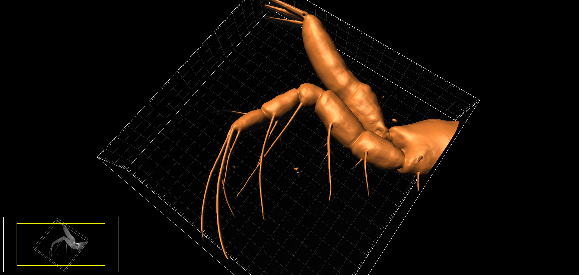
Drishti    Advanced software for 3D rendering of volumetric datasets.

FluoRender    Interactive 3D rendering tool for confocal microscopy designed specifically for neurobiologists.

Icy  open community platform for bioimage informatics. Broad selection of plugins and protocols.

ImageJ    Multi-format (Java-based) open source software package for data acquisition, analysis and processing. Extensive functionality conferred via a wide selection of downloadable plugins.

LAS-AF Lite    ‘Lite’ version of the Leica application suite which allows basic processing and analysis of  image data obtained from advanced Leica widefield and confocal systems.

LCS Lite    ‘Lite’ version of the Leica confocal software that allows basic processing and analysis of Leica SP2 confocal image files.

Micro-Manager    Open source software for control of automated microscopes which runs as a plugin to Image J.

Open Microscopy Environment (OMERO)    Client server software for visualisation, management and analysis of biological images.

DeconvolutionLab    Software for  deconvolution of 2D or 3D microscopic images which runs as a plugin to Image J.

V3D    Powerful open source software for visualisation and segmentation of large 3D datasets.

View5D    Software for analysis and processing of multi-dimensional volumetric datasets which runs as a plugin to Image J.

VisBio    Open source software for visualisation and analysis of multidimensional image data. Interfaces with Image J and OMERO.

Voxx    Voxel-based rendering software for 3D analysis of confocal and multi-photon datasets.

LSM image browser    Imaging software for Zeiss LSM 5 series confocals.

ZEN Lite    ‘Lite’ version of the Zeiss Efficient Navigation  (ZEN) software that allows basic processing and analysis of image data from advanced Zeiss light microscopical systems.

AJH

IN-FOCUS: Shedding light on new fluors: development of novel, metal-based probes for bioimaging.

Dr Simon Pope and his research group in CHEMY have been collaborating with the BIOSI Bioimaging Unit for over 8 years. Their research is focussed upon the development of new, metal-based, fluorescent probes for cell imaging applications, and forms part of a larger study on the use of metal complexes, which include rhenium and gold, as multi-modal imaging agents with therapeutic potential. In these studies, we have performed confocal imaging to (1) assess the cytotoxicity of the new probes, (2) evaluate their cellular uptake and determine their cytoplasmic localisation, and (3) characterise their fluorescent emissions via spectral (wavelength or lambda) scanning. The collaboration has yielded a number of high impact publications (see below) as well as a journal cover! With the improved potential of the new super-resolution confocal system we anticipate a lot more to come.

AJH

Find out more:

IN-FOCUS: De-boning the Zebrafish: unpicking skeletogenesis under the microscope.

Confocal reconstructions of the head, thorax and tail regions of the Zebrafish (Danio rerio)

Confocal reconstructions of the head, thorax and tail regions of the Zebrafish (Danio rerio)

The Zebrafish (Danio rerio) is, in many ways, the perfect model for microscopists. Not only does it share 70% genetic homology with man, but its larvae are born in large, transparent broods all year round and develop extremely quickly (a single cell develops into something resembling a fish within 24 hours!)  This means that developmental events can be visualised in vivo in real-time down the microscope. On top of this, their genome has been sequenced and it is easily amenable to molecular manipulation- again, these manipulations can be followed closely under the microscope lens.

Over the last few years we have been collaborating with Dr Chrissy Hammond at Bristol University, a fish biologist who shares an interest in skeletal development and disease. In our joint studies, we have used a variety of imaging techniques (brightfield, DIC, polarising, epifluorescence, confocal, TEM, radiography and microCT) to investigate skeletal development, growth and ageing in  this animal model.

One of the many interesting findings from our studies is that ageing fish undergo degenerative changes to their spine that resemble osteoarthritis (for example, spinal curvature, osteophyte formation, and connective tissue degeneration). This opens up the possibility  that they could be used to experimentally model aspects of the human disease. So it’s not just fishy tails!

AJH

Find out more:
Further Reading:

IN-FOCUS: Gumming up the works: oral biofilms under the microscope.

Image showing yeast cells colonising a tissue-engineered oral epithelium

Yeast cells (Candida albicans; red) colonise and invade a tissue-engineered oral epithelium (cell nuclei; blue; cytoplasm, green)

The BIOSI Bioimaging Facility has worked closely with Professor David Williams at the Dental School in Cardiff for a number of years. David is an expert in oral microbiology, specialising in microbial biofilms (e.g. dental plaque) and mycoses such as oral candidiases (thrush). Over this time, we have been involved in a number of collaborative studies where we have used confocal microscopy and various fluorescent labelling techniques to investigate the formation, 3D organization and microbial community structure of biofilms grown on tissue engineered oral epithelium, endotracheal tubes and substrates such as dental acrylic and titanium. The research has also evaluated the effect of various anti-microbial and anti-fungal compounds and commercial mouth rinses on biofilm development using fluorescent viability stains. The studies have extended our understanding of how oral biofilms develop and in how they respond to therapeutic intervention, and have resulted in a number of publications (see below) as well as a book cover for a leading text on the subject of Oral Microbiology. It’s a fantastic application of confocal microscopy to a biological problem and, from an imaging perspective, its been something for us to really get our teeth into!

AJH

Find out more:
Further reading:

NEWS: Naomi’s nightmares of nature.

Naomi

Image: BBC camera crew (left) filming the follicle mite, Demodex (right), for an episode of the children’s television programme, Naomi’s Nightmare’s of Nature.

Not so long ago we received a strange request from Dr Sarah Perkins (BIOSI): could we accommodate a BBC camera crew within the Bioimaging Facility to film an episode of the Children’s CBBC television programme, Naomi’s Nightmares of Nature? The nightmare in question, was the eyelash mite,  Demodex, a commensal ectoparasite that lives within the hair follicles (Demodex follicularum) and sebaceous glands (Demodex brevis) of the face, feeding off sebum and other organic detritus. Anyway, prior to filming, we spent an anxious morning attempting to isolate live Demodex from ‘volunteers’ faces by various means,  including via sellotape, with little success Fortunately, when Naomi and her production team arrived, we struck gold! A few eyelashes extracted from their cameraman, Steve, revealed a bumper load of parasites and, using DIC optics, we were able to generate some nice microscopic footage of a family of mites tucking into their evening meal!

AJH

Find out more:

IN-FOCUS: Heavy metal worms under the spotlight.

chloragocyte

Not just pretty pictures: an earthworm coelomocyte imaged by conventional confocal microscopy (left; DIC/fluorescence image) and via spectral scanning (centre) to analyse changes in riboflavin fluorescence caused by soil pollution (right).

The Research Techniques module run by Professor Pete Kille and Dr Carsten Muller (Introduction to Environmental Toxicology) makes for a busy week within the Bioimaging Research Hub. In the practical, students learn a range of advanced analytical research techniques as they aim to identify and characterize earthworm populations that have been sampled from land polluted by heavy metal – and I’m not talking about Axel’s Rose garden here : )

Pete is an expert in ecotoxicology and much of his research centers on how invertebrate species, such as the earthworm, deal with heavy metal pollutants, e.g., lead, in their environment. As it turns out, they seem to be pretty good at tolerating a lot of the nasty stuff that passes through them, but it does leave an indelible metabolic mark – making the organisms ideal for environmental toxicological testing. And here’s where it gets interesting: previous studies of the earthworm, Eisenia fetida, have shown that heavy metals affect riboflavin (vitamin B2) biosynthesis. Now, riboflavin happens to be (1) highly autofluorescent, and (2) neatly packaged within spheroidal organelles, or chloragosomes, within a sub-population of  immune cells, called coelomocytes, that are resident within the body cavity of the worm. Fortunately, earthworms can be gently persuaded to give up some of these cells for confocal microscopic analysis.

In the practical, we use confocal microscopy to image earthworm coelomocytes and, via spectral scanning, generate emission spectra of the riboflavin autofluorescence from within the chloragosomes. By comparing the autofluorescent signatures of coelomocytes from worms obtained from different sampling sites, we have asked the question: can riboflavin autofluorescence in this organism be used to assess soil pollution?

And the answer? Well, I’m not at liberty to say – the students reports aren’t in yet! (Answers on the back of a postcard to…)

AJH

Find out more:

Zimmerman et al. (2003) Spectral Imaging and its applications in live cell microscopy. FEBS Letters 546: 87-92.

Further Reading

IN-FOCUS: Imaging the Mitten crab: working hand in glove with the Natural History Museum

Imaris Snapshot

3D surface-rendered model of appendage (endopod of maxilliped) of Chinese mitten crab larva. Image produced on Zeiss LSM880 confocal and reconstructed using Bitplane Imaris.

Last week we undertook some confocal microscopy for the National History Museum to help characterise the arrangement of setae on the larval appendages of the Chinese mitten crab, Eriocheir sinesis * (now published, see Kamanli et al, 2017 below). The Mitten crab, so-named because of the tufts of ‘fur’ on the adult’s claws, is officially listed as one of the World’s most invasive species. The crabs out-compete and prey on native crab species, damage fishing nets and cause significant erosion of riverbanks, thus are of considerable economic importance.  They arrived in this country from China in the 1930’s via discharge of ballast water from trading ships and are now firmly established in many of Britain’s waterways. The National History Museum is investigating ways of reducing the population of Mitten crabs and this species is currently under evaluation as a potential food source in the UK (so if you can’t beat them, eat them!)

AJH

Find out more:
Further reading: