
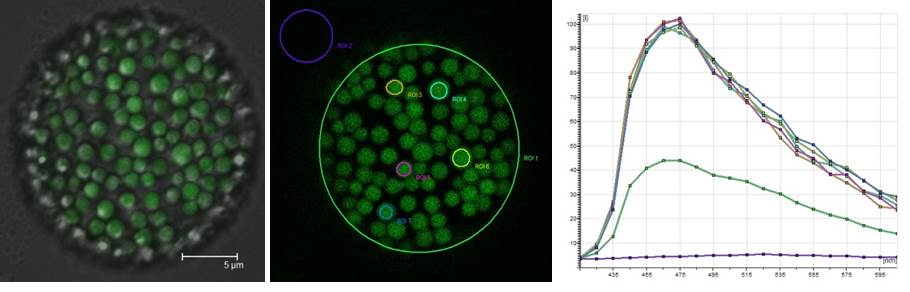
Not just pretty pictures: an earthworm coelomocyte imaged by conventional confocal microscopy (left; DIC/fluorescence image) and via spectral scanning (centre) to analyse changes in riboflavin fluorescence caused by soil pollution (right).
The Research Techniques module run by Professor Pete Kille and Dr Carsten Muller (Introduction to Environmental Toxicology) makes for a busy week within the Bioimaging Research Hub. In the practical, students learn a range of advanced analytical research techniques as they aim to identify and characterize earthworm populations that have been sampled from land polluted by heavy metal – and I’m not talking about Axel’s Rose garden here : )
Pete is an expert in ecotoxicology and much of his research centers on how invertebrate species, such as the earthworm, deal with heavy metal pollutants, e.g., lead, in their environment. As it turns out, they seem to be pretty good at tolerating a lot of the nasty stuff that passes through them, but it does leave an indelible metabolic mark – making the organisms ideal for environmental toxicological testing. And here’s where it gets interesting: previous studies of the earthworm, Eisenia fetida, have shown that heavy metals affect riboflavin (vitamin B2) biosynthesis. Now, riboflavin happens to be (1) highly autofluorescent, and (2) neatly packaged within spheroidal organelles, or chloragosomes, within a sub-population of immune cells, called coelomocytes, that are resident within the body cavity of the worm. Fortunately, earthworms can be gently persuaded to give up some of these cells for confocal microscopic analysis.
In the practical, we use confocal microscopy to image earthworm coelomocytes and, via spectral scanning, generate emission spectra of the riboflavin autofluorescence from within the chloragosomes. By comparing the autofluorescent signatures of coelomocytes from worms obtained from different sampling sites, we have asked the question: can riboflavin autofluorescence in this organism be used to assess soil pollution?
And the answer? Well, I’m not at liberty to say – the students reports aren’t in yet! (Answers on the back of a postcard to…)
AJH
Find out more:
Zimmerman et al. (2003) Spectral Imaging and its applications in live cell microscopy. FEBS Letters 546: 87-92.
Further Reading
- Koziol et al. (2006) Riboflavin as a source of autofluorescence in Eisenia fetida coelomocytes. Photochem Photobiol 82: 570-573
- Plytycz et al. (2009) Riboflavin content of coelomocytes in earthworms (Dendrodrilus rubidus) field populations as a molecular biomarker of soil metal pollution. Environ Pollut 157: 3042-3050