Featured post

IN FOCUS: The ‘3D Pollen Library’: An Update

Above: Transparent 3D rendering of a dandelion (Taraxacum officinale) pollen grain. Surface exine displayed in green, inner intine structure in blue. Image produced by Dr Anthony Hayes, Bioimaging Hub, Cardiff School of Biosciences.

Over the last 12 months I’ve been working closely with biovisualisation specialist Dr Kristen Brown at NIH3D to curate our 3D pollen model resources into a purpose-built 3D collection:  the ‘3D Pollen Library’. This collection is now featured on the NIH3D homepage and, at the time of writing, represents the largest collection of 3D pollen grain models worldwide. To date, it contains over a hundred entries together with taxonomic metadata and links to other well-established online pollen resources – a significant achievement considering its humble beginnings (you can read more about the background work leading up to it here.)

Enormous thanks must go to Kristen and her team who have been incredibly helpful throughout in accommodating my (many) requests regarding the ‘look and feel’ of the curated collection. They have done an absolutely splendid job.

Above: A small selection of surface rendered pollen grains in the 3D Pollen Library collection. These examples were recently created in collaboration with Dr Heather Pardoe, Amgueddfa Cymru, National Museum Wales.

Above: the user interface for visualisation and manipulation of the 3D pollen models. The models are all viewable  as surface rendered or wireframe meshes and can be downloaded in x3d, stl, glb and wrl file formats for 3D printing and AR/VR visualisation. The source confocal data is also available for download, as is the published methodology for creating the models.

In parallel with the above, I’ve established an ongoing collaboration with Dr Heather Pardoe (senior botanist and chief palynologist at Amgueddfa Cymru, National Museum Wales). This collaboration has allowed us to digitise and 3D model pollen grains and spores from selected plant species in the museum’s extensive archival pollen collection using methodology we’ve developed in-house at the Bioimaging Hub. The 3D pollen models produced via this collaboration will be added to our NIH3D library as a separate pollen sub-collection, as well as being viewable as part of the full collection. It is envisaged that these models will have significant utility as educational tools for teaching and exhibition.

Further reading:


IN FOCUS: Breaking the Mould: Supersizing Pollen Grains as Gigantic Public Art Sculptures.

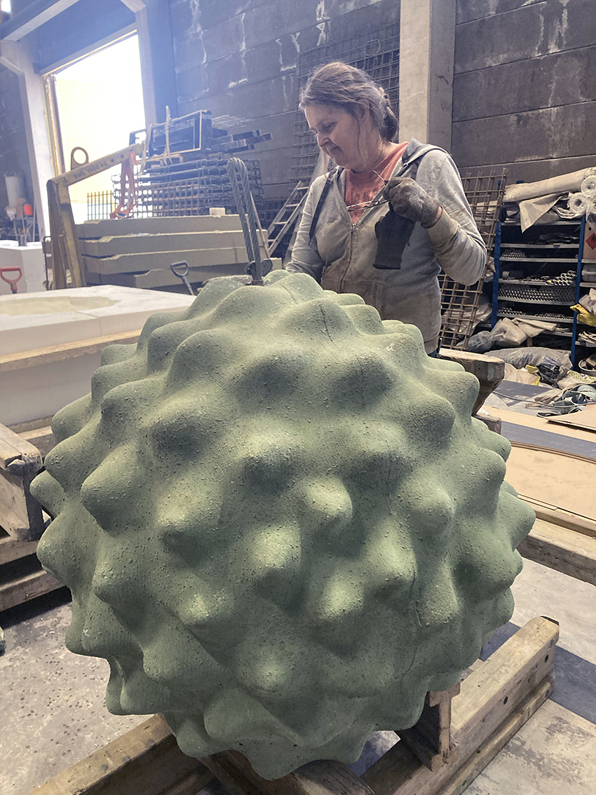
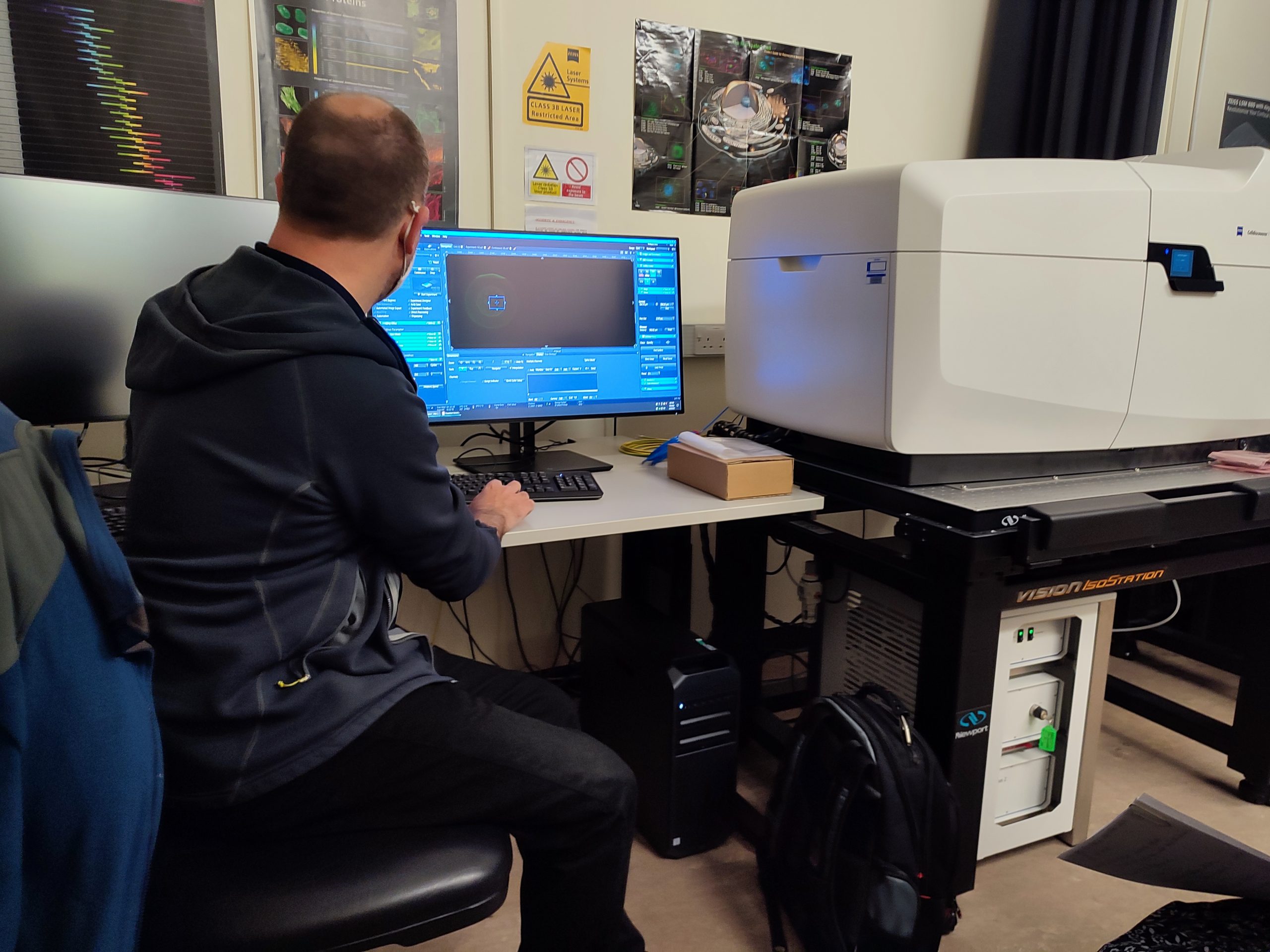
Above: A pollen grain from ragweed supersized in eco-friendly natural concrete. The concrete sculpture is one metre in diameter, weighing approximately one and a quarter tonne  – about the same weight as a small car! For comparison, its ‘little brother’, i.e. the source pollen grain it was based upon, was approximately twenty microns in diameter, thus enlarged by a factor of 500,000x. Photo courtesy of Saara Ekström.

You’d be forgiven for thinking that a microscope would be essential to explore the intricate 3D structure of a pollen grain. Well, how about climbing on top of it and exploring it by hand instead? Confused? Please read on.

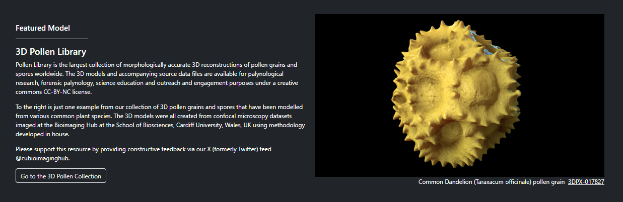
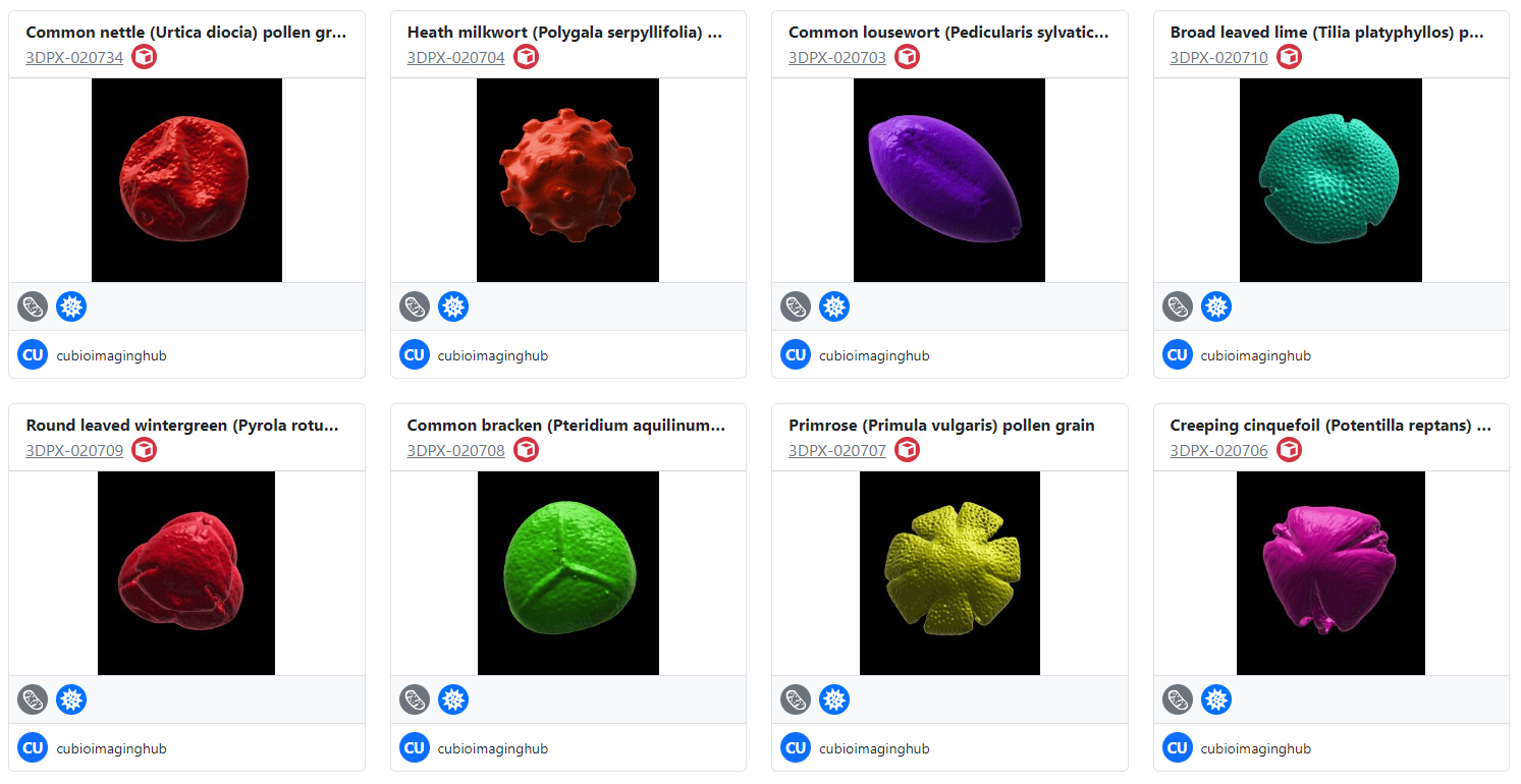
If you’ve visited this site before then, chances are, you’ll be aware that I enjoy ‘noodling around with pollen’ : )  Over the last seven years,  the Bioimaging hub has developed  and refined methodology that permits  3D printing and both virtual and augmented reality (VR and AR, respectively) visualization of microscopic samples such as pollen grains for use in educational and immersive learning experiences. A direct offshoot of this work has been the development of  what is  currently the largest online repository of 3D pollen grain models worldwide.  To date, I have curated over 170 published, DOI-referenced 3D pollen models from over 150 species of plant which can be downloaded free, for non-commercial usage, via  the Bioimaging Hub’s 3D Pollen Library Collection on the NIH3D website You can read more about the evolution of this resource in the links below (including a recent article I have written for the Royal Microscopical Society In Focus magazine, December edition – to follow).

Recently, the project took another interesting and unexpected evolutionary turn. I was contacted by Finnish visual artist and film maker, Saara Ekström, who pitched to me an amazing idea for a collaboration: gigantifying some of our 3D pollen models as public art sculptures for an elementary school in Helsinki, Finland. The plan was to cast the sculptures in coloured eco-friendly natural concrete at a range of sizes between 0.7  and 2 metres to create a magical ‘Alice in Wonderland’ outdoor learning environment for the children, helping to raise their awareness of the unseen, natural world and fostering an understanding of ‘one of nature’s smallest and most essential components’. An absolutely wonderful idea! 

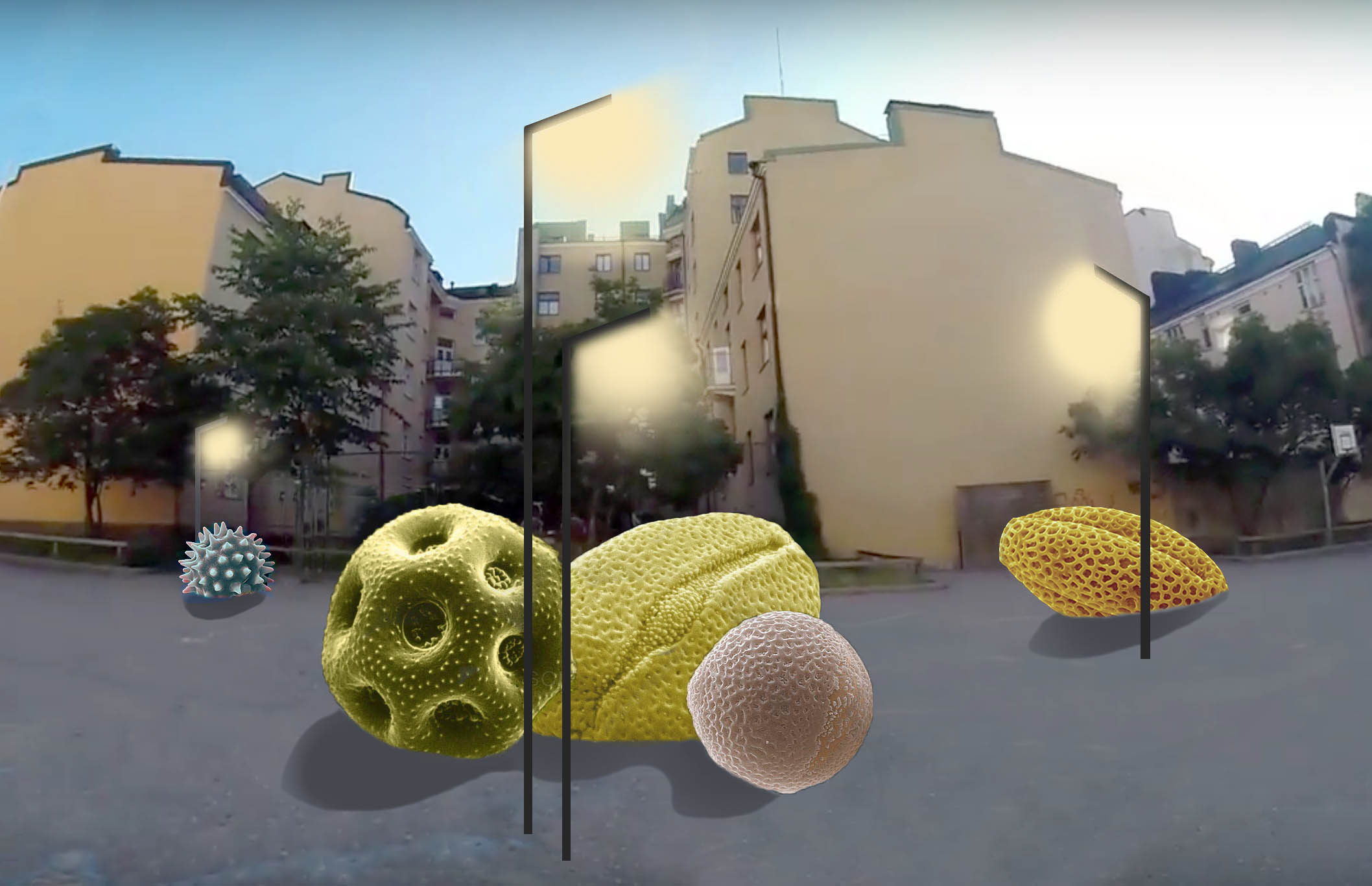
Above: Concept art for the 3D pollen sculptures courtesy of Saara Ekström.

As part of the project, a protected piece of old-growth forest, purchased from the Finnish Natural Heritage Foundation, will be presented as a gift to the School, thus preserving something that is vitally important to future generations of children and linking the pollen sculptures to this natural ecosystem. Having kids of my own (and ever mindful of the ongoing environmental crisis) I was delighted to support Saara’s ambitious project.  The thought that pollen grains from flowers in my back garden would be reproduced as two metre concrete megaliths nearly two thousand miles away in Helsinki was also simply irresistible! Stonehenge, nothing ; )

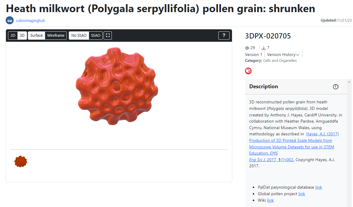
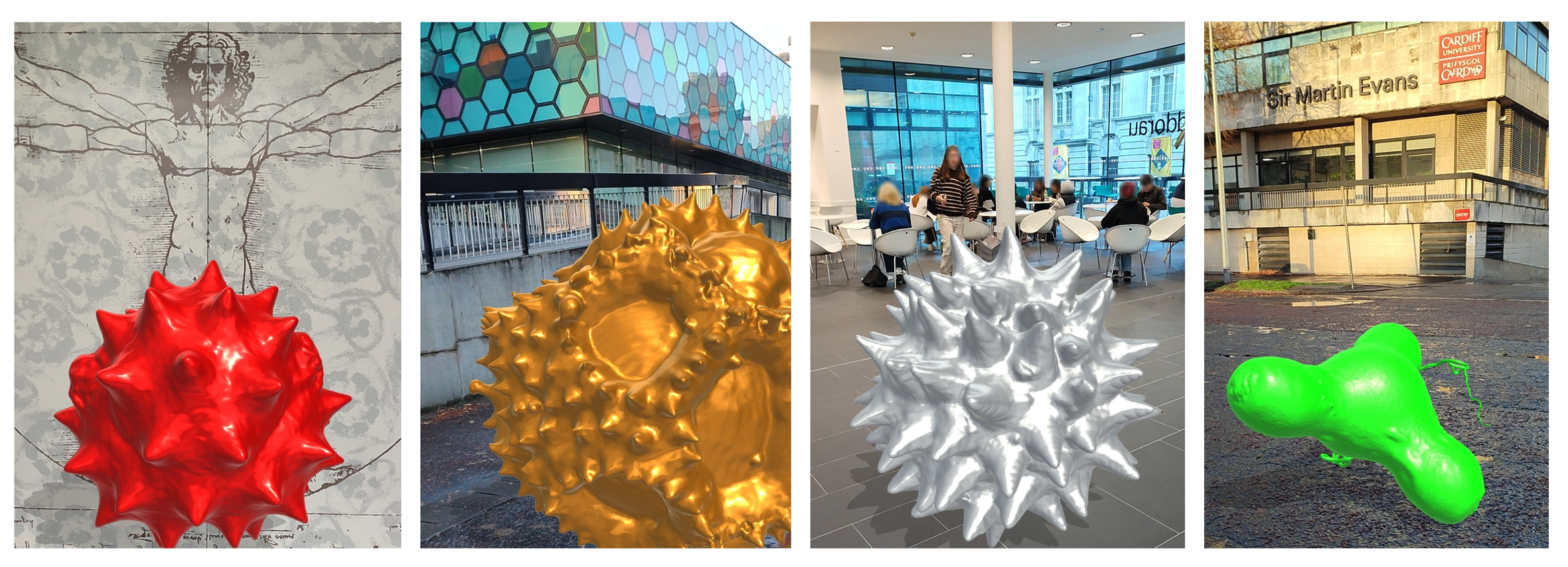
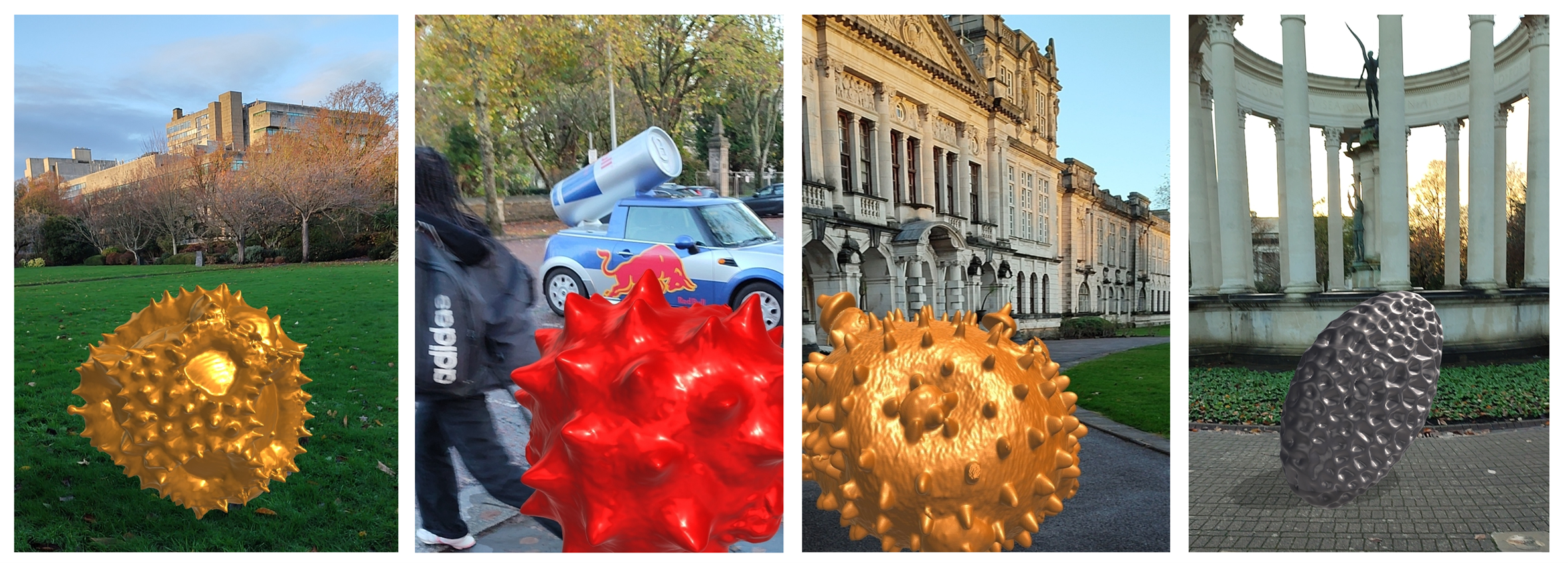
Saara identified four pollen species of  interest from our collection: blue passion flower (Passiflora caerulea); common thyme (Thymus vulgaris); black elderberry (Sambucus nigra) and common ragweed (Ambrosia sp.which exhibited strikingly different morphologies and surface ornamentation (see below). Some of the more spiky and ornate pollen grain species (e.g. common daisy, Bellis perennis) in our collection were deliberately avoided since the children would have to be able to climb on them safely without risk of impalement!

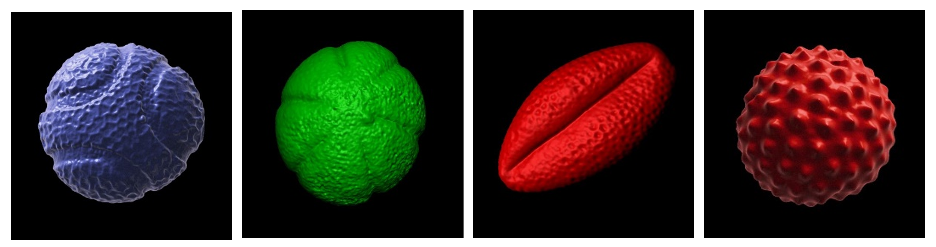
Above: the four species of pollen grain to be supersized as concrete sculptures. From left to right: blue passion flower; common thyme; black elderberry and common ragweed. 3D pollen models created by Tony Hayes. The 3D models are available for download for non-commercial usage from the Bioimaging Hub’s 3D Pollen Library collection at NIH3D.

So how does one create a gigantic concrete sculpture of a microscopic pollen grain? Well, this was one of the first things I asked Saara, who was more than happy to explain the steps involved. First, our 3D pollen meshes are downloaded from the NIH3D website and scaled up into virtual models of the sculptures using CAD software, taking care to retain as much surface detail as possible (see below). The pollen grain mesh is also truncated in order to form a flat, stable base (we wouldn’t want them to accidentally roll onto someone’s foot, after all!)

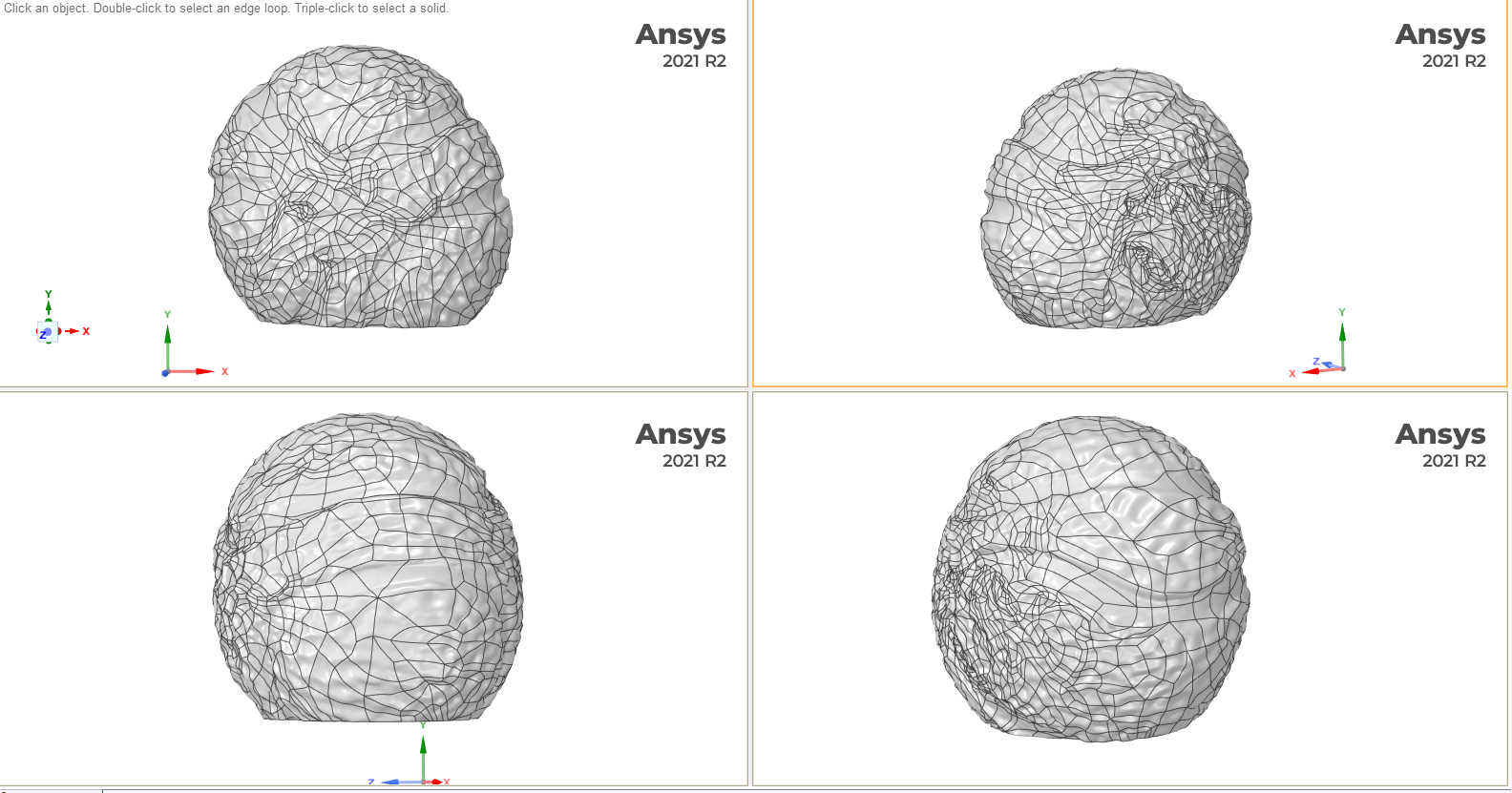
Above: 3D meshes of the pollen grain (Blue passion flower shown here in multiple orientations) are used to program a tool that mechanically carves a negative form into styrofoam thereby forming the casting moulds. Screen grabs courtesy of Saara Ekström.

The meshes are then used to program a tool that mechanically carves a negative form of the 3D models into large, high density styrofoam blocks to create the complementary halves of the casting moulds, as shown below.

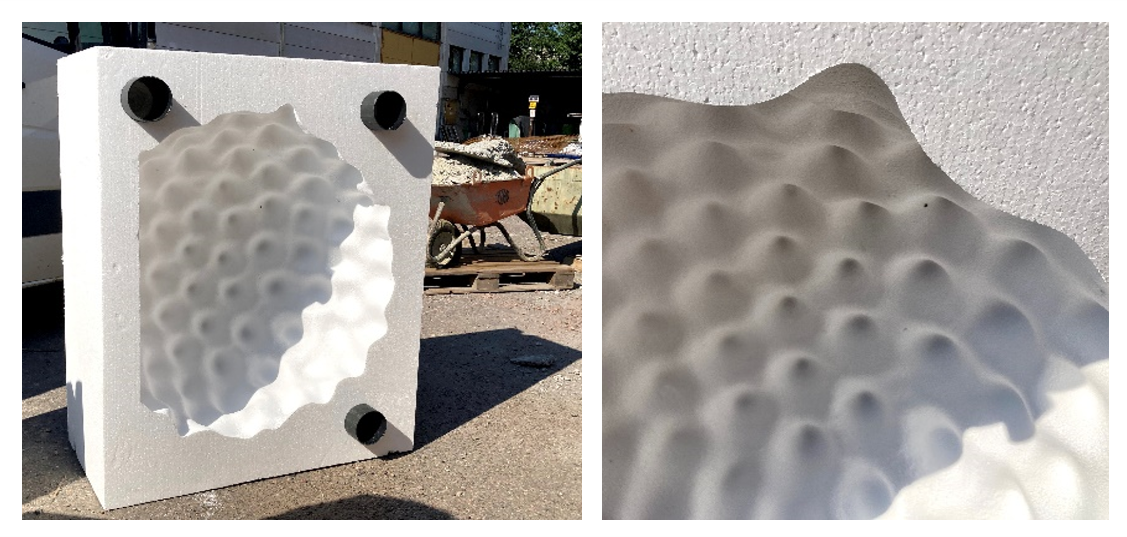
Above: The casting moulds (Ragweed pollen shown here) are mechanically carved out of high density styrofoam blocks and are able to record surface detail with high fidelity. A flat base has been introduced into the mould (left panel) to maintain stability of the resultant sculpture. The protruding black cylinders allow the two complementary halves of the mould to be held tightly together during the casting process.  A wheel barrow in the background demonstrates the large scale of the moulds. Photos courtesy of Saara Ekström.

Once the moulds are created, steel reinforcement is incorporated into the cavity to add structural strength to the sculpture. Coloured eco-friendly natural concrete is then cast, using different coloured pigments for each pollen species. Once the concrete has cured, the moulds are removed and (in the words of Saara) ‘Volia, a microscopic pollen grain has grown to nearly monumental proportions!’ 

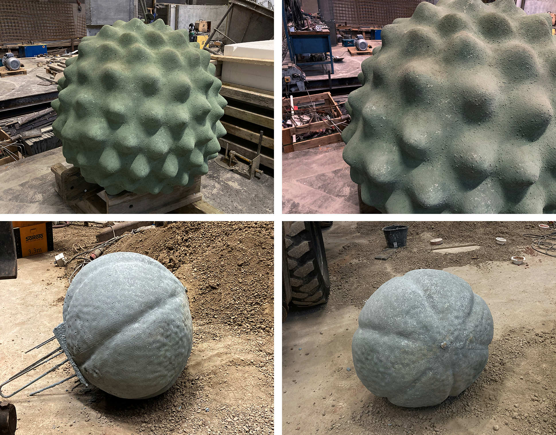
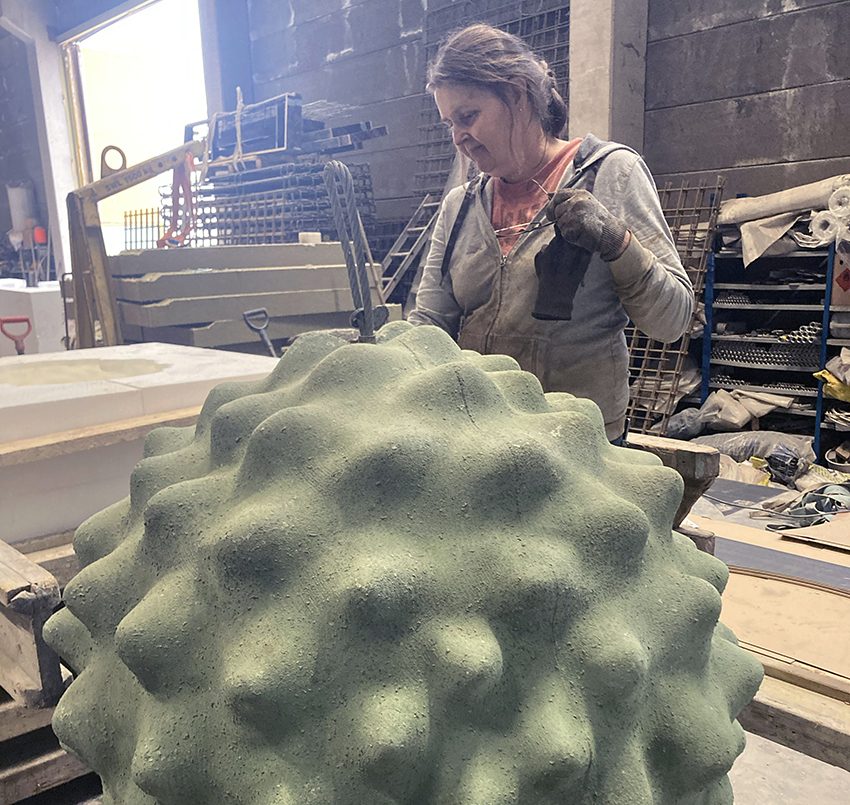
Above: The unfinished concrete pollen sculptures following removal of the styrofoam moulds. Top panel shows Ragweed pollen, bottom panel shows Thyme pollen. Rebar steel reinforcement can be seen protruding from the base of the thyme pollen sculpture (bottom left). Photos courtesy of Saara Ekström.

The concrete sculptures are then hand finished and a coloured protective coating is added before they are moved to their final destination, the Kallio primary school in Helsinki. At the site they are carefully lowered into place by crane. Rubber protective matting is then installed around each sculpture.

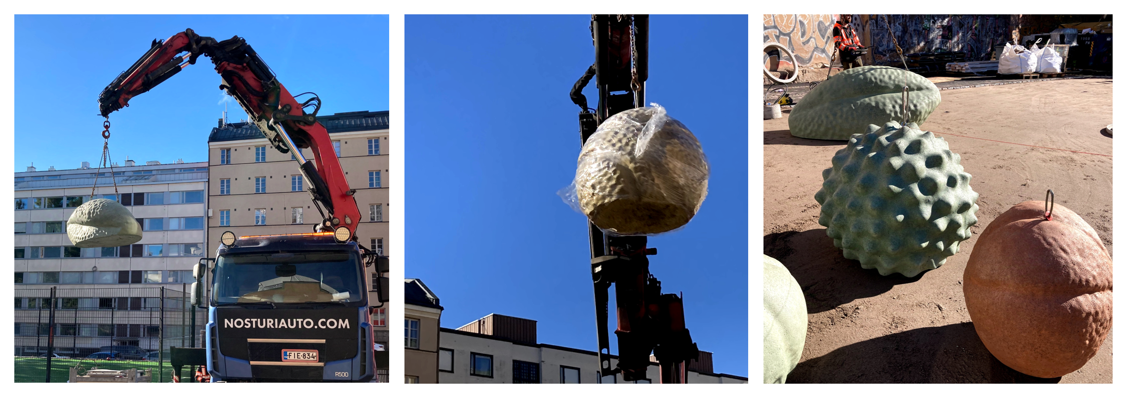
Above: A crane lowers the concrete sculptures into place at the Kallio primary school, Helsinki. Left panel, black elderberry pollen sculpture; centre panel, passionflower pollen sculpture; right panel, some of the concrete sculptures in situ. The precise placement of the sculptures is specified in the artists design. Photos courtesy of Saara Ekström.

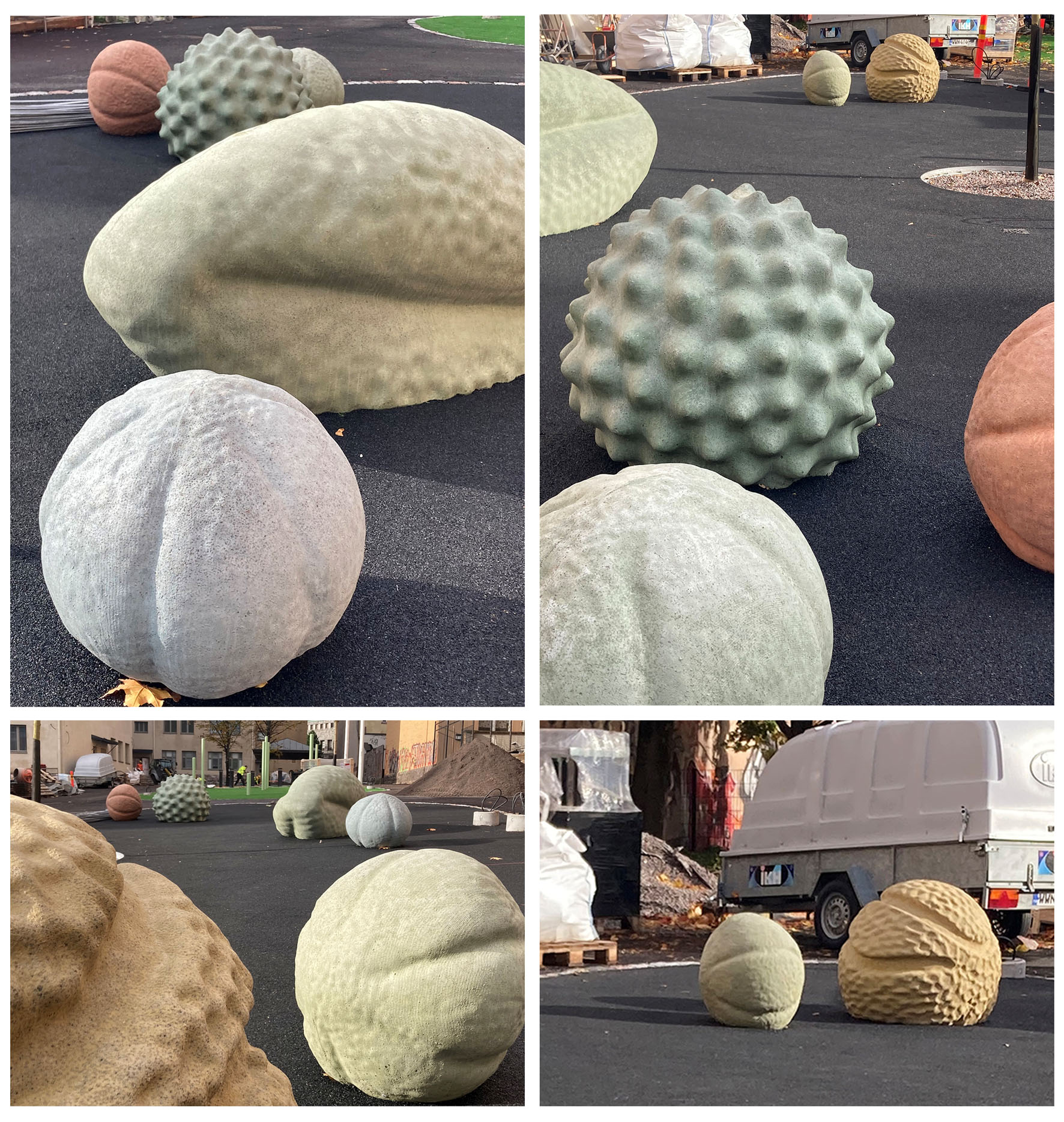
Above: The finished pollen sculptures surrounded by rubber safety matting. There are seven sculptures in total in a range of different colours: four of the thyme pollen and one each of the other three  species,. The area will be landscaped with trees and mood lighting to create an inviting ambience. Photos courtesy of Saara Ekström.

As the winters are long and dark in Finland, customised mood lighting will illuminate the sculptures creating a warm and inviting ambience, as depicted in the concept art above. I think it is fair to say that Saara, has done an absolutely monumental job!

The pollen sculptures were commissioned by the City of Helsinki and will be administered by the Helsinki Art Museum who are responsible for all the city’s public art collection.

AJH 25/09/2024

Further reading


IN FOCUS: Making the Most of Your Microscope.

Above: A selection of stage plate inserts 3D printed by the Bioimaging Research Hub – links to resources in blog article below.

Hands up if your microscope is badly in need of upgrade or repair but your budget won’t stretch that far? Maybe a new focusing knob to replace the one that just broke off in your hand, or perhaps a new stage plate adapter, reflector cube or filter holder to increase your imaging options? Perhaps a C-mount or smartphone adaptor to give one of your old microscopes a new lease of life? Or even a sample holder or chamber for a bespoke imaging application? What the heck, let’s think big eh? How about a completely new modular microscope system with tile scanning capabilities?

Way too expensive, eh?… Well, imagine for a moment that you could just click a button (or a few buttons, at least) and make it so. If you haven’t yet realised, I’m talking about 3D printing in light microscopy and the life sciences – the subject of a very interesting paper that I recently came across  – see below.

It’s safe to say that 3D printing is changing the way we do things in microscopy, now permitting low-cost upgrade, repair, or customisation of microscopes like never before. There are now a huge selection of 3D printable resources available through websites such as NIH3D, Thingiverse etc that can be used to modify your microscope system or to generate scientific apparatus or labware for upstream sample processing and preparation procedures.

So, to save you trawling through the 3D printing sites in order to identify the most useful designs to meet your histology and imaging needs we’ve done it for you and have curated a list of 3D printable resources which we hope you’ll find useful (below).

AJH 25/05/2023

Further Reading

—————————————————————-

3D printable resources for histology and light microscopy. Information collated by Dr Tony Hayes, Bioimaging Research Hub, School of Biosciences, Cardiff University, Wales, UK.

 Sample processing

Sample staining

Sample storage and archiving

 Sample presentation

Microscope: Complete builds

Microscope: Maintenance

Microscope: Phone adapters (a selection)

Microscope: Stands (a selection)

Microscope: Components (generic)

Microscope: Olympus-specific

 Microscopes: Leica-specific

Microscopes: Zeiss-specific

 Microscopes: Nikon-specific

IN FOCUS: AR Palynology: Probing the Reality of Nature/Nature of Reality.

Above: Augmented reality visualisation of pollen grains 3D rendered in gigantic proportions. You wouldn’t want one of these getting stuck up your nose!

Back in 2015 we developed some novel methodology that allowed us to generate 3D printed models from confocal z-stacks of microscopic samples.  At the time, we showcased the technique by making 3D prints of pollen grains from various plant species for use in science education and outreach activities, musing that a physical, tangible model would allow improved 3D conceptualization of these microscopic structures – original blog article here. The work generated a fair amount of interest and resulted in requests for 3D printed models of pollen grains from far and wide (e.g., the Smithsonian National Museum of Natural History in the US, the UK Met Office, National Botanical Garden of Wales etc). Following publication of our methodology in 2017 other researchers soon followed suit, creating their own 3D printed models from confocal datasets utilizing methods similar to ours. By this time, we had turned our attention to virtual reality (VR) as a tool to experience/manipulate microscopic objects, as well as larger more complex biological models in virtual space – something we saw as a logical  evolution of the 3D learning experience. You can read more about this in a separate blog and see the progress we made in developing the resource via a short video on our YouTube site.

Fast-forward to present, a global pandemic, accelerating environmental change, and a cost-of-living crisis  – the economic fallout from COVID, the folly of Brexit, a war in Ukraine, and gross government incompetence. To borrow a quote from a certain Vladimir Ilyich Ulyanov, there are decades where nothing happens, and there are weeks where decades happen. Well, quite!

But what of the 3D pollen models? Well, for one, coronaphobia has meant that people are less inclined these days to be handling and passing around shared resources such as plastic pollen models or VR headsets at risk of spreading/contracting germs. Moreover, plastic is increasingly being viewed negatively due to the environmental damage caused by plastics and ‘frivolous’ 3D printing could be seen as increasing the existing plastic problem. Lastly, with the cost-of-living crisis/looming recession, it would seem that no one can afford to do anything other than feed themselves and try to keep warm these days, let alone commission 3D pollen prints from the Bioimaging Hub – particularly since there’s now a growing number of pollen models available through 3D printing sites such as Sketchfab etc. And so, with these concerns in mind, we’ve donned our thinking caps and come up with a plan to address the prevailing zeitgeist.

Firstly, we’ve decided to make our entire repository of 3D pollen models publicly available, free of charge, under a Creative Commons CC-NY-NC licence via the newly developed NIH3D website (formerly the NIH3D print exchange) which is a leading  community-driven portal for sharing and downloading  bioscientific content for 3D printing and interactive 3D visualization. Under the terms of our licence, the models can be used, shared, or modified for non-commercial purposes, as long as the creator(s) are properly credited. To date, we’ve made available 3D models of seventy distinct species of pollen grains and spores through the Bioimaging Hub’s new NIH 3D profile page which have now been curated into a special collection, the 3D Pollen Library We have also included all source data (confocal z-stack files in Carl Zeiss Image .czi file format). To the best of our knowledge, this represents the largest single collection of 3D pollen files available online and the plan is to add more models plus supporting data in future. For cross-referencing, relevant links have been included to major palynological databases, PalDat and the Global Pollen project, and, of course,  Wikipedia – the fount of all knowledge : ) We’ve also showcased a small selection of our pollen models on the Bioimaging Hub’s Sketchfab site – these can’t be downloaded directly, however you can manipulate the models on screen and view them in VR – further information here.

Secondly, we’ve been experimenting with augmented reality (AR) as a tool to allow visualization and exploration of our 3D models in real-world environments. The beauty of AR, of course, is that it doesn’t require a headset,  just a smartphone or tablet which are now more ubiquitous than ever.  Thus, a 3D model in the relevant file format  (usually  .glb or .gltf) can be downloaded directly to an individual’s smartphone or tablet and, using appropriate AR software, seamlessly integrated with real time digital information from the camera for display on the touchscreen. This allows users to personally experience a real environment with generated perceptual information overlaid upon it. Within the AR environment, the models can be scaled up or down, freely moved and rotated via the touchscreen, or circumnavigated and explored both externally and internally via directional information from the device’s sensors, à la Pokemon-GO. This allows for a  highly realistic and immersive interactive experience (different from the artificial environments of VR) thus facilitating 3D conceptualization of the embedded model. Furthermore, freed from the physical encumbrance of a VR headset, the user is less likely to blunder into office furniture, moving traffic etc or experience the nausea, headaches and dizziness of VR-associated cybersickness.

So how does one view our 3D pollen models in AR? Well, it’s quite straightforward really, but to make things even easier I’ve put together a set of instructions (below) that should get you up and running in next to no time:

  1. Install a free AR app on your mobile device (smartphone or tablet) from the Google Play or Apple App stores that handles .glb (GL transmission format binary) files – this is the common standard file format for AR/VR visualisation.  There are quite a lot of AR apps to choose from and we’ve only tested the NeoSpace AR app on android devices but this seems to work quite well.
  2. Go to the Bioimaging Hub’s NIH 3D webpage – link here – select a 3D pollen model, and then click on the DOWNLOAD option. You will then see a list of  the files that we’ve made available for download (refer to screenshot below). These include:
  •  .glb file format for AR/VR applications (available as both zipped and unzipped files).
  • .stl (stereolithography, .wrl (‘worlds’ virtual reality modelling) and x3d file formats for 3D printing applications.
  • source confocal data in .czi (Carl Zeiss Image) file format (zipped file).

  1. For AR viewing, download the .glb file to your mobile device (if zipped then extract the compressed file).
  2. Open the .glb file in the AR viewer app and follow the on-screen instructions. Initially you’ll have to scan your environment with the camera on your smartphone/tablet so that the app can identify a flat surface in order to place the 3D model into your display.

If you’ve done it correctly then you should now be able to view any of our 3D pollen models in whatever context your imagination, mobile device and AR app allows. Below are some examples  of the type of AR imagery that we’ve generated using a free android AR app.

Above:  Video sequence captured from an  android smartphone running the free AR app, NeoSpace.  The 3D pollen model can be zoomed, freely moved and rotated via touchscreen and also circumnavigated and explored both externally and internally via directional information from the device’s sensors.

Above: Attack of the giant pollen grains! Photo-realistic AR imagery generated using an android smartphone and the NeoSpace AR app.

We’d love to get some feedback with photos or videos showing how you have used our pollen models for 3D printing, or AR/VR applications in science education and outreach. Please keep us updated via our twitter account @cubioimaginghub. Best of luck!

AJH, November, 2022 (updated March 2023).


Further reading:

NEWS: New BIOSI Live Cell Imaging Spoke

Above: Spinning disc confocal microscope set up in the new BIOSI live cell imaging spoke

A dedicated live  cell imaging spoke has been set up in the Sir Martin Evans Building (BIOSI; E/3.15). The microscopy suite has inverted widefield, scanning and spinning disc confocal microscopes with full environmental control systems. Further information available through Pete Watson.

AJH 08/04/2022


CORE EQUIPMENT: New Zeiss Celldiscoverer 7 system

Above: Training on the Bioimaging Hub’s new Zeiss Celldiscover7 imaging system

The Bioimaging Hub has recently taken delivery of a state-of-the-art, automated  live cell imaging system to replace its old Leica SP2 confocal microscope. The Celldiscoverer7 imaging system, which was purchased via the generous support of Cardiff University’s Research Infrastructure Fund (Lead Applicant: Dr Tony Hayes), has the latest Zeiss LSM 900 confocal scan head with Airyscan 2 detector technology and is capable of multi-format, high-throughput and super-resolution analysis of a wide range of samples from cell cultures to small model organisms. The system supports photomanipulation via FRAP, FRET and related techniques and is furnished with a comprehensive Zen software package that includes modules for deconvolution and machine learning, amongst other cutting edge features. Further details of the system are available through the Bioimaging Hub’s research equipment database.

AJH

Find out more:

NEWS: Updated Covid Rules: Resumption of Hands-on Support and Training.

A huge and heartfelt thank you to all users and support staff of the Bioimaging Hub for your strict adherence to our covid security measures over the last 12 months. It has been an extremely difficult year for all of us and we have tried to manage the situation as effectively and as safely as possible, working within the security framework provided by Cardiff University and Welsh government.

In line with the latest guidance, we are pleased to now begin relaxing some of our covid security measures and to be in a position where we can reintroduce direct hands-on support and training for our microscope systems.  To facilitate this provision, it remains vitally important that users follow the new guidance protocols, as detailed below.

Before entering the Bioimaging Hub:

  • All users must familiarize themselves with current Welsh Government Coronavirus (Covid-19) Guidance and read the Bioimaging Hub’s updated Coronavirus risk assessment.
  • Users must not visit the Bioimaging Hub if they are displaying any symptoms of Covid-19; if they have been in a high risk area; or had recent contact with a covid-positive individual without confirmation of a negative test result.
  • All users are advised to make regular use of the Cardiff University covid screening service  or to utilise rapid flow testing methodology that is now widely available through the NHS and pharmacies.

Room occupancy status and technical support/training:

  • Room occupancy status has now increased to two independent users/two research bubbles per microscopy suite, up to a maximum of four people per microscopy suite in total.
  • Direct hands-on training and support for our microscope systems will now resume under social distancing rules and with PPE including face coverings.

COVID working Regulations within the Bioimaging Research Hub:

  • Users should continue to use the room booking calendars and should specify which microscope system they require in the title section of the booking request (i.e., specify user name and microscope system). Booking details here.
  • Social distancing rules remain in place within all areas of the Bioimaging Hub.
  • PPE remains mandatory including the use of face coverings in all communal areas.
  • Microscope cleaning procedures before and after use remain mandatory.
  • Histology sample drop-offs and collections should continue to be arranged via email (bioimaginghub@cardiff.ac.uk).

Thanks.

AJH 28.9.2021

IN-FOCUS: Brushing Up On Your Background Knowledge.

With the dizzying pace of technological innovation and ongoing advances in microscopy and imaging, it is becoming increasingly difficult to keep abreast current developments in the field. With this in mind, we’ve rounded up some essential resources, from basic to advanced, that will keep you informed and updated.

Online magazines & journals:

Educational portals:

AJH

SPOKE EQUIPMENT: Olympus VS200 High Throughput Slide Scanning Microscope.

Above: the new Olympus VS200 high throughput slide scanning system in ECSCRI

A new, high throughput slide scanning system has recently been installed in the European Cancer Stem Cell Research Institute (ECSCRI) and is available for use as a spoke of the Bioimaging Research Hub. The equipment allows automated high-throughput scanning of histological samples via a range of image modalities, including epifluorescence. Further details of the system are available through the Hub’s equipment database. All enquiries for this system should be directed towards Mr Mark Bishop.

Further Reading:

AJH

NEWS: Reopening of the Bioimaging Research Hub: New COVID 19-Security Measures.

For the time being, use of the Bioimaging Hub is going to be a very different experience for everyone. In drawing up these guidelines we’ve taken a lead from the Welsh Government and have started with a very cautious approach that we’ll constantly review and relax where possible. For now, the guidance may seem quite stringent so please bear with us while we all adapt to this new way of working.

Access is not permitted to the Bioimaging Research Hub without (i) completing a risk assessment form that should cover all planned activities within the facility and (ii) reading all relevant supporting information (see below). A template risk assessment is available through the Bioimaging Hub’s main web pages.

Before completing the risk assessment form, all users must download and read the following documents:

Cardiff University’s COVID-19 secure organisational risk assessment

The Bioimaging Hub’s local COVID-19 security guidelines

DO NOT enter the Bioimaging Research Hub:

  • If you are SARS-Cov-2 positive or are displaying symptoms of COVID-19 infection (e.g. persistent coughing, elevated temperature, anosmia, sickness).
  • If you have been in a high-risk area or have had recent contact with confirmed SARS-CoV-2 positive individuals within the last 14 days.
  • If you have an underlying health condition and are concerned it will put you at greater risk of developing severe COVID-19 symptoms. N.B. essential work can be undertaken by Bioimaging Hub staff at a supported rate if necessary.

COVID Working Regulations within the Bioimaging Research Hub

All users of the facility:

  • MUST read the above documentation before entering the Bioimaging Research Hub.
  • MUST contact bioimaginghub@cardiff.ac.uk before entering the facility.You are not permitted to simply drop-in unplanned. Drop off/collection of histology samples must be arranged in advance. Researchers must knock before entering histology via E/0.08 and observe all safety directives outlined in this document. Users must complete a histology request form to specify processing preferences in advance of their visit.
  • MUST wash their hands upon entering and leaving the facility (hand wash station opposite office area). Multiple gel hand wash dispensers are also available within the microscopy suites.
  • MUST wear appropriate personal protective equipment (PPE) within the facility, including a buttoned-up lab coat and gloves (gloves are provided in all microscopy suites). Eye protection is advisable.
  • MUST clean the imaging equipment and immediate working area before and after use (cleaning instructions are available at each microscope station; alcohol spray and lab roll have been provided for this purpose).
  • MUST observe social distancing (i.e. 2 metres inter-personal space) within the Hub. The main corridor should be used by only one person at a time. While social distancing measures are in place no training is available and technical support will be provided on a remote basis via telephone (main office: 02920876611; shared office: 02922510220; histology: 02920875139).
  • MUST NOT enter the staff office area (0.14A) or histology suites (E/0.06-E/0.07). Drop off/collection of histology samples must be arranged in advance (use E/0.08).
  • MUST knock before entering any room. All microscopy suites are now single occupancy (i.e. one in, one out with at least 15 mins user-free time between bookings). The microscope booking calendars have been replaced with room booking calendars, as follows: “BIOSI – E/0.03 – Confocal/Lightsheet microscopy”, “BIOSI – E/0.04 – widefield microscopy”, “BIOSI – E/0.05 – spinning disc microscopy”. New booking instructions can be found here.
  • MUST set the room occupancy status (vacant/in use) on the door sliders of the microscopy suites before and after use.

FURTHER READING

German BioImaging recommendations for operating Imaging Core Facilities in a research environment during the SARS-CoV-2 pandemic

Leica: How to sanitize a microscope

Olympus: How to clean and sterilize your microscope

Zeiss: Cleaning and disinfecting the microscope and its optical components