
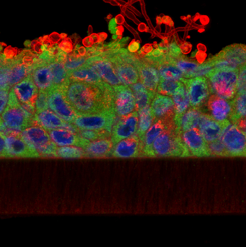
Yeast cells (Candida albicans; red) colonise and invade a tissue-engineered oral epithelium (cell nuclei; blue; cytoplasm, green)
The BIOSI Bioimaging Facility has worked closely with Professor David Williams at the Dental School in Cardiff for a number of years. David is an expert in oral microbiology, specialising in microbial biofilms (e.g. dental plaque) and mycoses such as oral candidiases (thrush). Over this time, we have been involved in a number of collaborative studies where we have used confocal microscopy and various fluorescent labelling techniques to investigate the formation, 3D organization and microbial community structure of biofilms grown on tissue engineered oral epithelium, endotracheal tubes and substrates such as dental acrylic and titanium. The research has also evaluated the effect of various anti-microbial and anti-fungal compounds and commercial mouth rinses on biofilm development using fluorescent viability stains. The studies have extended our understanding of how oral biofilms develop and in how they respond to therapeutic intervention, and have resulted in a number of publications (see below) as well as a book cover for a leading text on the subject of Oral Microbiology. It’s a fantastic application of confocal microscopy to a biological problem and, from an imaging perspective, its been something for us to really get our teeth into!
AJH
Find out more:
- Neu et al. (2014) Investigation of microbial film structure by laser scanning microscopy. Productive Biofilms 146: 1-51
- Palmer &Sternberg (1999) Modern microscopy in biofilm research: confocal microscopy and other approaches. Curr Opin Biotechnol 10: 263-268
Further reading:
- Cavalcanti et al. (2015) Virulence and pathogenicity of Candida albicans is enhanced in biofilms containing oral bacteria. Biofouling 31: 27-28
- Boros-Majewska et al. (2014) Novel Nystatin A1 derivatives exhibiting low host cell toxicity and antifungal activity in an in vitro model of oral candidosis. Medical Microbiology and Immunology 203: 341-355
- Hooper et al. (2012) The visualisation and speed of kill of wound isolates on a silver alginate dressing. International Wound Journal 9: 633-642
- Silva, et al. (2010) Candida glabrata and Candida albicans co-infection of an in vitro oral epithelium J Oral Pathol Med 50: 421-427
- Malic et al. (2009) Detection and identification of specific bacteria in wound biofilms using peptide nucleic acid (PNA) fluorescent in situ hybridisation (PNA FISH) Microbiol 155: 2603-2611
- Malic et al (2006) Characterisation of Candida albicans infection of an in vitro oral epithelial model using confocal laser scanning microscopy. Oral Microbiol Immunol 22: 188-194