
THE 3D POLLEN LIBRARY: from humble beginnings to the largest online collection of morphologically-accurate 3D printable pollen models.
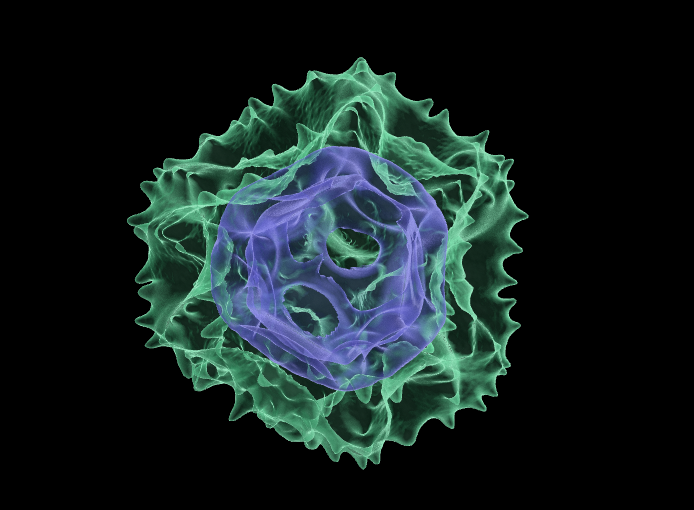
Above: Transparent 3D rendering of a dandelion (Taraxacum sp) pollen grain showing outer sculpted exine (green) and internal cage-like intine (blue) – just one of over seventy pollen species represented in the Bioimaging Hub’s 3D pollen collection.
Back in 2015, the bioimaging hub developed some novel methodology allowing us to 3D print scale models of microscopic samples from volume datasets obtained via optical sectioning microscopy (i.e., confocal, lightsheet, multiphoton microscopy etc). The 3D outputs generated via this procedure are morphologically accurate and highly scalable making them extremely useful tactile learning resources for science education, outreach and exhibition purposes.
Following on from this work, I wrote a series of short blog articles (links below) where I showcased the technique by 3D printing scale models of microscopic pollen grains (see below), utilising the autofluorescence from the outer wall, or exine, of the pollen grain to fabricate the 3D print. In 2017, I wrote up and published the work as a methods paper (link below) allowing other researchers to employ the technique to make 3D models of microscopic structures for use in their research, teaching and science outreach/engagement activities. Surprisingly, both the blogs and the paper generated a fair amount of interest at local, national and even international level, and we received a number of requests asking us to create 3D pollen prints and even curated collections for a number of science establishments (examples below). We therefore continued to add new pollen species to our growing repository whenever time permitted. By 2019, we had turned our attention to VR and had begun experimenting with this sensory modality as a way of physically manipulating our 3D digital models (both anatomical and microscopic) in virtual space using the Oculus series VR headsets and controllers, later developing workflows for their augmented reality visualisation. You can read more about these developments in the blogs below.
We have now made the decision to make our entire back-catalogue of 3D pollen models available free of charge for non-commercial usage through the new NIH 3D site (formerly the NIH 3D Print Exchange) under a Creative Commons CC-BY-NC licence. The 3D models and all accompanying source data (Carl Zeiss Image .czi files) are available for download for palynological research, paleoarcheology, forensic palynology, science education and outreach and engagement purposes through the Bioimaging Hub’s 3D Pollen library. The included file formats support 3D printing and augmented reality and virtual reality visualisation. For each entry, links are provided to the PalDat palynological database, Global pollen project and also Wikipedia.
Bioimaging Hub 3D Pollen Library (all models) through NIH 3D
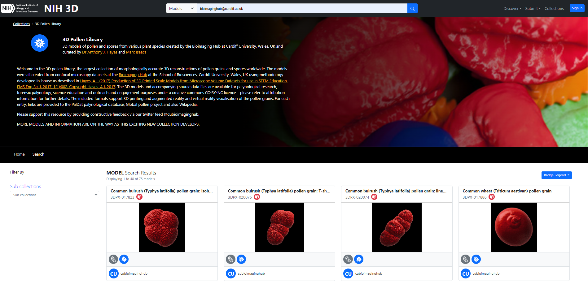
Above: homepage of the Bioimaging Hub’s 3D Pollen Library collection.
We have also showcased some of our 3D pollen models on the Bioimaging Hub’s Sketchfab site (see below). These can’t be downloaded directly, however you can manipulate the models on screen, customise lighting, textures etc and also view them in VR (further details below).
Bioimaging Hub 3D pollen collection through Sketchfab
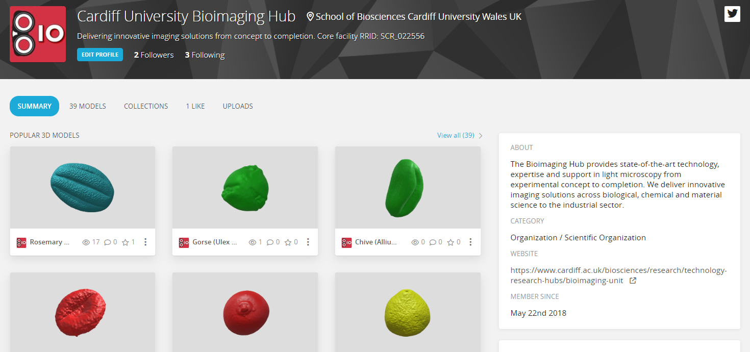
Above: Homepage of the Bioimaging Hub’s Sketchfab 3D pollen collection.
Please feel free to peruse and download some (or all) of our 3D models. We’d welcome constructive feedback with photographs via our twitter feed @cubioimaginghub. This will allow us to continue to develop and improve these resources.
AJH 29/03/23
Further reading:
- Perry, I., Szeto, J-Y., Isaacs, M.D., Gealy, E.C., Rose, R., Scofield, S., Watson, P.D., Hayes, A.J. (2017) Production of 3D printed scale models from microscope volume datasets for use in STEM education. EMS Engineering Science Journal. 1 (1): 002
- IN FOCUS: Bigging it up: 3D printing to change the shape of microscopy (February, 2016).
- IN FOCUS: Development of a 3D printed pollen reference collection (August, 2016)
- IN FOCUS: 3D Pollen prints not to be sniffed at: printing pollen for the met office (July, 2017).
- IN FOCUS: Immersive Microscopy: 3D visualisation and manipulation of microscopic samples through virtual reality (February, 2019)
- IN FOCUS: Plastic fantastic: making pollen models for the National Botanic Garden of Wales.(October, 2019)
- IN FOCUS: AR Palynology: probing the reality of nature/nature of reality (November, 2022)