
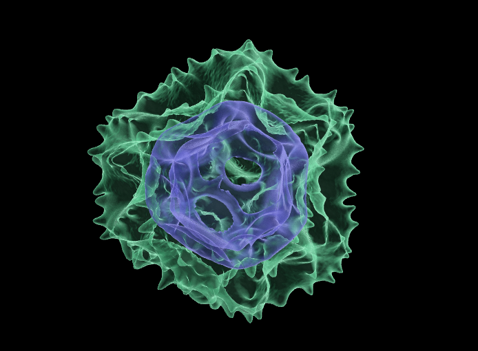
Above: Transparent 3D rendering of a dandelion (Taraxacum officinale) pollen grain. Surface exine displayed in green, inner intine structure in blue. Image produced by Dr Anthony Hayes, Bioimaging Hub, Cardiff School of Biosciences.
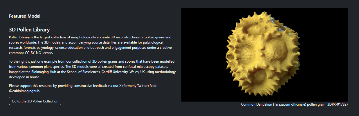
Over the last 12 months I’ve been working closely with biovisualisation specialist Dr Kristen Brown at NIH3D to curate our 3D pollen model resources into a purpose-built 3D collection: the ‘3D Pollen Library’. This collection is now featured on the NIH3D homepage and, at the time of writing, represents the largest collection of 3D pollen grain models worldwide. To date, it contains over a hundred entries together with taxonomic metadata and links to other well-established online pollen resources – a significant achievement considering its humble beginnings (you can read more about the background work leading up to it here.)
Enormous thanks must go to Kristen and her team who have been incredibly helpful throughout in accommodating my (many) requests regarding the ‘look and feel’ of the curated collection. They have done an absolutely splendid job.
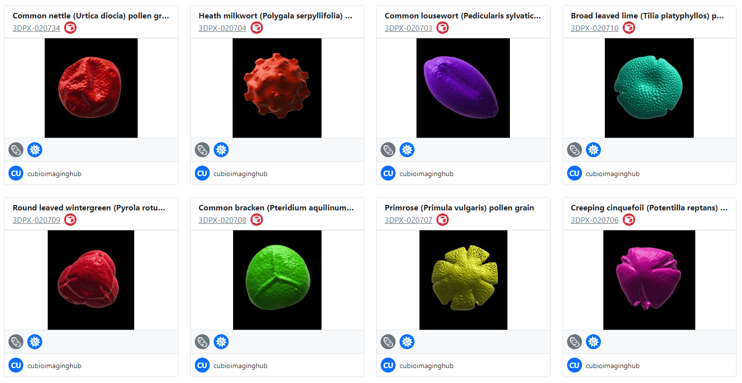
Above: A small selection of surface rendered pollen grains in the 3D Pollen Library collection. These examples were recently created in collaboration with Dr Heather Pardoe, Amgueddfa Cymru, National Museum Wales.
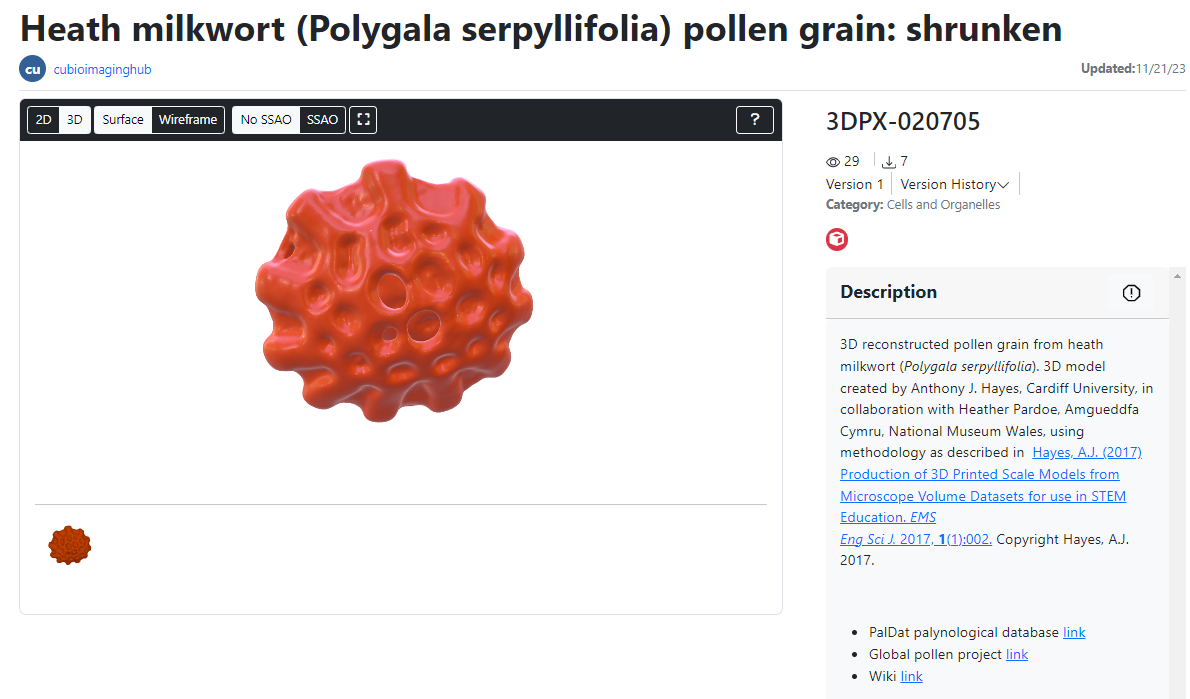
Above: the user interface for visualisation and manipulation of the 3D pollen models. The models are all viewable as surface rendered or wireframe meshes and can be downloaded in x3d, stl, glb and wrl file formats for 3D printing and AR/VR visualisation. The source confocal data is also available for download, as is the published methodology for creating the models.
In parallel with the above, I’ve established an ongoing collaboration with Dr Heather Pardoe (senior botanist and chief palynologist at Amgueddfa Cymru, National Museum Wales). This collaboration has allowed us to digitise and 3D model pollen grains and spores from selected plant species in the museum’s extensive archival pollen collection using methodology we’ve developed in-house at the Bioimaging Hub. The 3D pollen models produced via this collaboration will be added to our NIH3D library as a separate pollen sub-collection, as well as being viewable as part of the full collection. It is envisaged that these models will have significant utility as educational tools for teaching and exhibition.
Please feel free to visit the 3D Pollen Library and download some of our models (they’re all free!) We’d welcome constructive feedback with photographs via our X (twitter) feed @cubioimaginghub which will allow us to continue developing and improving the 3D resource.
AJH 11/01/2024
Further reading:
- Perry, I., Szeto, J-Y., Isaacs, M.D., Gealy, E.C., Rose, R., Scofield, S., Watson, P.D., Hayes, A.J. (2017) Production of 3D printed scale models from microscope volume datasets for use in STEM education. EMS Engineering Science Journal. 1 (1): 002
- IN FOCUS: Bigging it up: 3D printing to change the shape of microscopy (February, 2016).
- IN FOCUS: Development of a 3D printed pollen reference collection (August, 2016)
- IN FOCUS: 3D Pollen prints not to be sniffed at: printing pollen for the met office (July, 2017).
- IN FOCUS: Immersive Microscopy: 3D visualisation and manipulation of microscopic samples through virtual reality (February, 2019)
- IN FOCUS: Plastic fantastic: making pollen models for the National Botanic Garden of Wales.(October, 2019)
- IN FOCUS: AR Palynology: probing the reality of nature/nature of reality (November, 2022)
- THE 3D POLLEN LIBRARY: from humble beginnings to the largest online collection of morphologically-accurate 3D printable pollen models.