
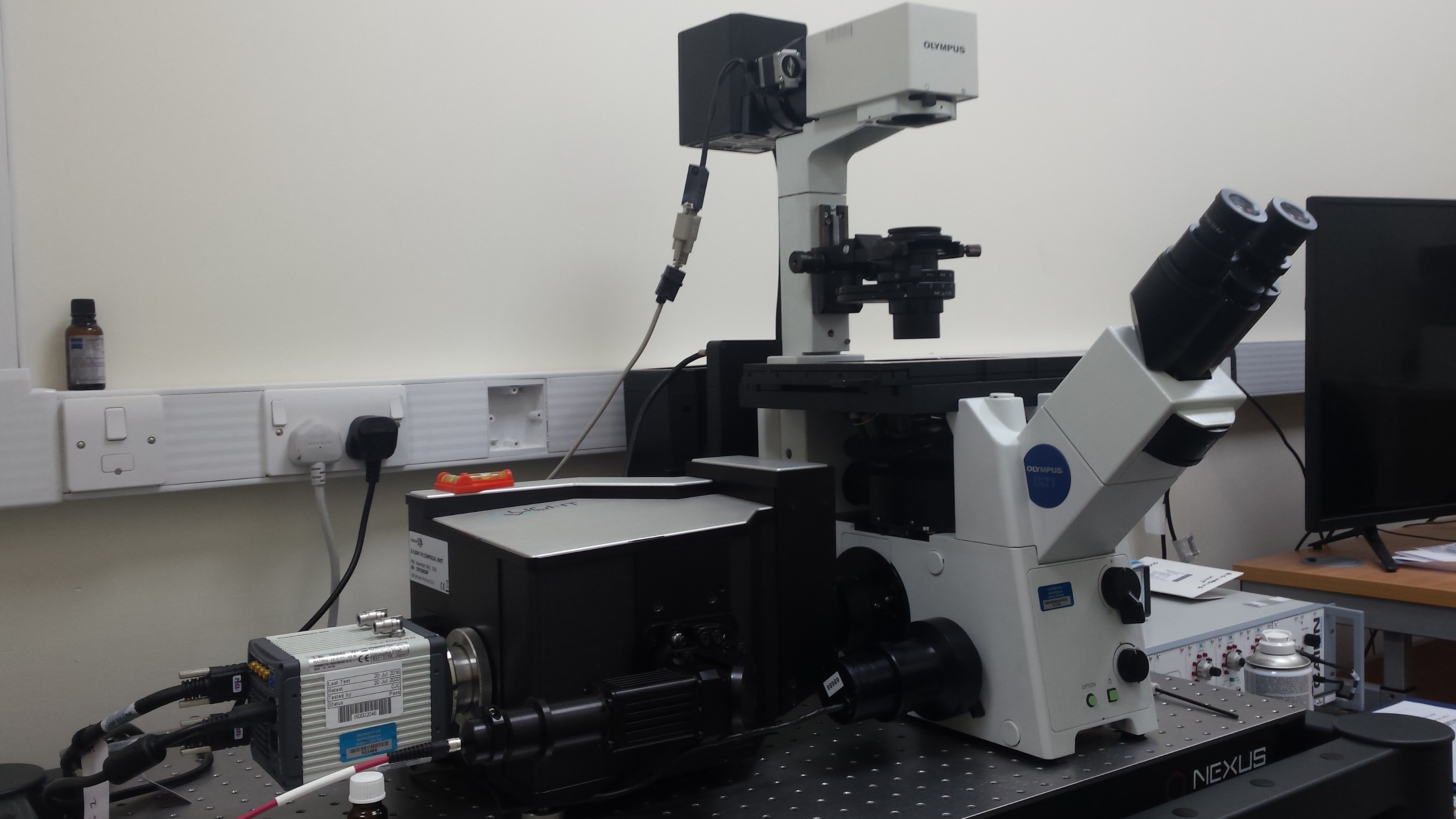
Above: The new spinning disc confocal system
The old electron microscopy darkroom (BIOSI 2; E/0.05) within the Bioimaging Research Hub has recently been refurbished as a live cell imaging suite via generous support from BIOSI. It now houses a spinning disc confocal system for fast, live cell imaging applications. The system is based around an Olympus IX71 inverted microscope, kindly provided by Dr Pete Watson, which has been upgraded, via ISSF funding, with a Crest Optics X-Light V2 confocal head, a Cairn Research tri-line laser bank (405nm, 488nm, 561nm) and a Hamamatsu ORCA Flash 4 sCMOS digital camera with M-View Gemini image splitter. The system is fully integrated via Molecular Devices MetaMorph software and boasts a 40″ 4k display. The system will expand the Hub’s imaging toolbox, enabling high speed, multi-position, multi-colour 3D/4D image acquisition. Support systems for live cell imaging (i.e. gas and incubation) are also available within the facility. Further details of this system are available through our equipment database.
AJH