
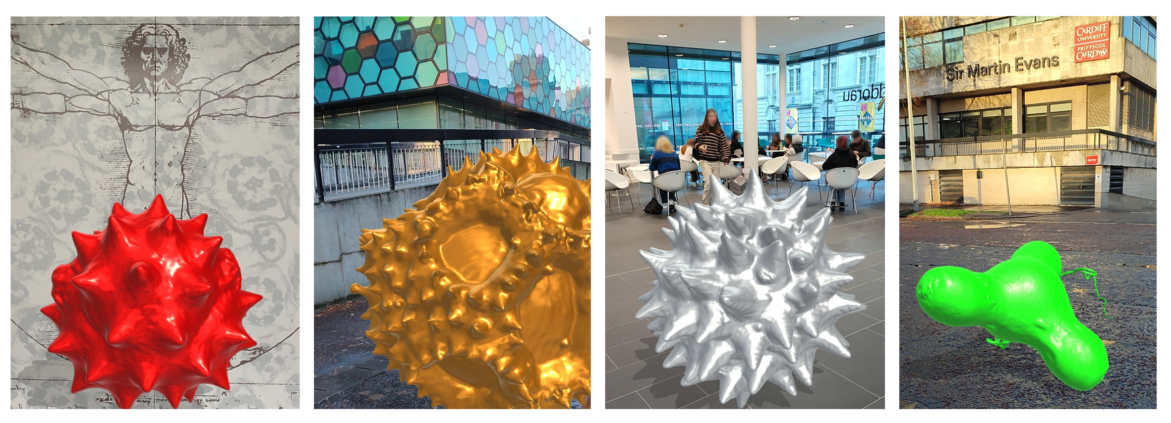
Above: Augmented reality visualisation of pollen grains 3D rendered in gigantic proportions. You wouldn’t want one of these getting stuck up your nose!
Back in 2015 we developed some novel methodology that allowed us to generate 3D printed models from confocal z-stacks of microscopic samples. At the time, we showcased the technique by making 3D prints of pollen grains from various plant species for use in science education and outreach activities, musing that a physical, tangible model would allow improved 3D conceptualization of these microscopic structures – original blog article here. The work generated a fair amount of interest and resulted in requests for 3D printed models of pollen grains from far and wide (e.g., the Smithsonian National Museum of Natural History in the US, the UK Met Office, National Botanical Garden of Wales etc). Following publication of our methodology in 2017 other researchers soon followed suit, creating their own 3D printed models from confocal datasets utilizing methods similar to ours. By this time, we had turned our attention to virtual reality (VR) as a tool to experience/manipulate microscopic objects, as well as larger more complex biological models in virtual space – something we saw as a logical evolution of the 3D learning experience. You can read more about this in a separate blog and see the progress we made in developing the resource via a short video on our YouTube site.
Fast-forward to present, a global pandemic, accelerating environmental change, and a cost-of-living crisis – the economic fallout from COVID, the folly of Brexit, a war in Ukraine, and gross government incompetence. To borrow a quote from a certain Vladimir Ilyich Ulyanov, there are decades where nothing happens, and there are weeks where decades happen. Well, quite!
But what of the 3D pollen models? Well, for one, coronaphobia has meant that people are less inclined these days to be handling and passing around shared resources such as plastic pollen models or VR headsets at risk of spreading/contracting germs. Moreover, plastic is increasingly being viewed negatively due to the environmental damage caused by plastics and ‘frivolous’ 3D printing could be seen as increasing the existing plastic problem. Lastly, with the cost-of-living crisis/looming recession, it would seem that no one can afford to do anything other than feed themselves and try to keep warm these days, let alone commission 3D pollen prints from the Bioimaging Hub – particularly since there’s now a growing number of pollen models available through 3D printing sites such as Sketchfab etc. And so, with these concerns in mind, we’ve donned our thinking caps and come up with a plan to address the prevailing zeitgeist.
Firstly, we’ve decided to make our entire repository of 3D pollen models publicly available, free of charge, under a Creative Commons CC-NY-NC licence via the newly developed NIH3D website (formerly the NIH3D print exchange) which is a leading community-driven portal for sharing and downloading bioscientific content for 3D printing and interactive 3D visualization. Under the terms of our licence, the models can be used, shared, or modified for non-commercial purposes, as long as the creator(s) are properly credited. To date, we’ve made available 3D models of seventy distinct species of pollen grains and spores through the Bioimaging Hub’s new NIH 3D profile page which have now been curated into a special collection, the 3D Pollen Library We have also included all source data (confocal z-stack files in Carl Zeiss Image .czi file format). To the best of our knowledge, this represents the largest single collection of 3D pollen files available online and the plan is to add more models plus supporting data in future. For cross-referencing, relevant links have been included to major palynological databases, PalDat and the Global Pollen project, and, of course, Wikipedia – the fount of all knowledge : ) We’ve also showcased a small selection of our pollen models on the Bioimaging Hub’s Sketchfab site – these can’t be downloaded directly, however you can manipulate the models on screen and view them in VR – further information here.
Secondly, we’ve been experimenting with augmented reality (AR) as a tool to allow visualization and exploration of our 3D models in real-world environments. The beauty of AR, of course, is that it doesn’t require a headset, just a smartphone or tablet which are now more ubiquitous than ever. Thus, a 3D model in the relevant file format (usually .glb or .gltf) can be downloaded directly to an individual’s smartphone or tablet and, using appropriate AR software, seamlessly integrated with real time digital information from the camera for display on the touchscreen. This allows users to personally experience a real environment with generated perceptual information overlaid upon it. Within the AR environment, the models can be scaled up or down, freely moved and rotated via the touchscreen, or circumnavigated and explored both externally and internally via directional information from the device’s sensors, à la Pokemon-GO. This allows for a highly realistic and immersive interactive experience (different from the artificial environments of VR) thus facilitating 3D conceptualization of the embedded model. Furthermore, freed from the physical encumbrance of a VR headset, the user is less likely to blunder into office furniture, moving traffic etc or experience the nausea, headaches and dizziness of VR-associated cybersickness.
So how does one view our 3D pollen models in AR? Well, it’s quite straightforward really, but to make things even easier I’ve put together a set of instructions (below) that should get you up and running in next to no time:
- Install a free AR app on your mobile device (smartphone or tablet) from the Google Play or Apple App stores that handles .glb (GL transmission format binary) files – this is the common standard file format for AR/VR visualisation. There are quite a lot of AR apps to choose from and we’ve only tested the NeoSpace AR app on android devices but this seems to work quite well.
- Go to the Bioimaging Hub’s NIH 3D webpage – link here – select a 3D pollen model, and then click on the DOWNLOAD option. You will then see a list of the files that we’ve made available for download (refer to screenshot below). These include:
- .glb file format for AR/VR applications (available as both zipped and unzipped files).
- .stl (stereolithography, .wrl (‘worlds’ virtual reality modelling) and x3d file formats for 3D printing applications.
- source confocal data in .czi (Carl Zeiss Image) file format (zipped file).

- For AR viewing, download the .glb file to your mobile device (if zipped then extract the compressed file).
- Open the .glb file in the AR viewer app and follow the on-screen instructions. Initially you’ll have to scan your environment with the camera on your smartphone/tablet so that the app can identify a flat surface in order to place the 3D model into your display.
If you’ve done it correctly then you should now be able to view any of our 3D pollen models in whatever context your imagination, mobile device and AR app allows. Below are some examples of the type of AR imagery that we’ve generated using a free android AR app.
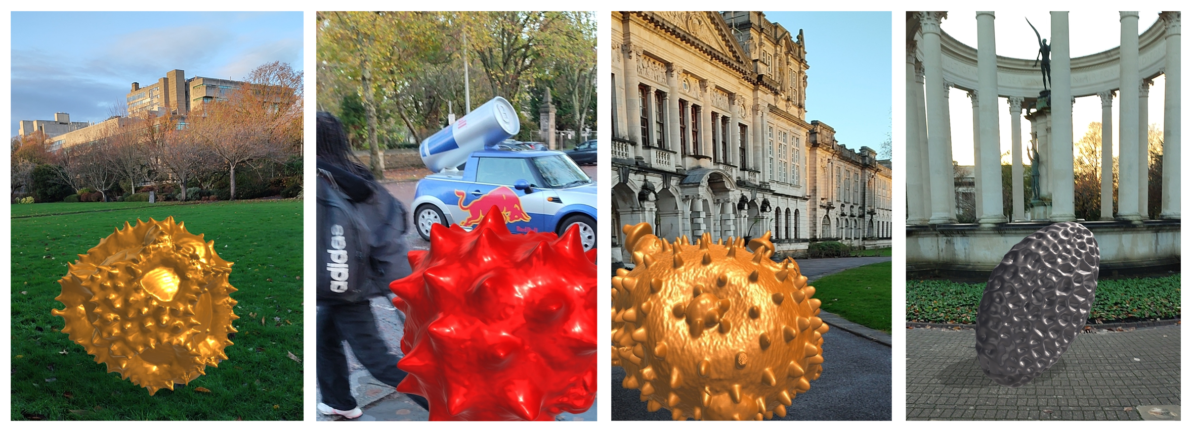
Above: Attack of the giant pollen grains! Photo-realistic AR imagery generated using an android smartphone and the NeoSpace AR app.
We’d love to get some feedback with photos or videos showing how you have used our pollen models for 3D printing, or AR/VR applications in science education and outreach. Please keep us updated via our twitter account @cubioimaginghub. Best of luck!
AJH, November, 2022 (updated March 2023).
Further reading:
- Perry, I., Szeto, J-Y., Isaacs, M.D., Gealy, E.C., Rose, R., Scofield, S., Watson, P.D., Hayes, A.J. (2017) Production of 3D printed scale models from microscope volume datasets for use in STEM education. EMS Engineering Science Journal. 1 (1): 002
- IN FOCUS: Bigging it up: 3D printing to change the shape of microscopy (February, 2016).
- IN FOCUS: Development of a 3D printed pollen reference collection (August, 2016)
- IN FOCUS: 3D Pollen prints not to be sniffed at: printing pollen for the met office (July, 2017).
- IN FOCUS: Immersive Microscopy: 3D visualisation and manipulation of microscopic samples through virtual reality (February, 2019)
- IN FOCUS: Plastic fantastic: making pollen models for the National Botanic Garden of Wales.(October, 2019)
- IN FOCUS: AR Palynology: probing the reality of nature/nature of reality (November, 2022)