
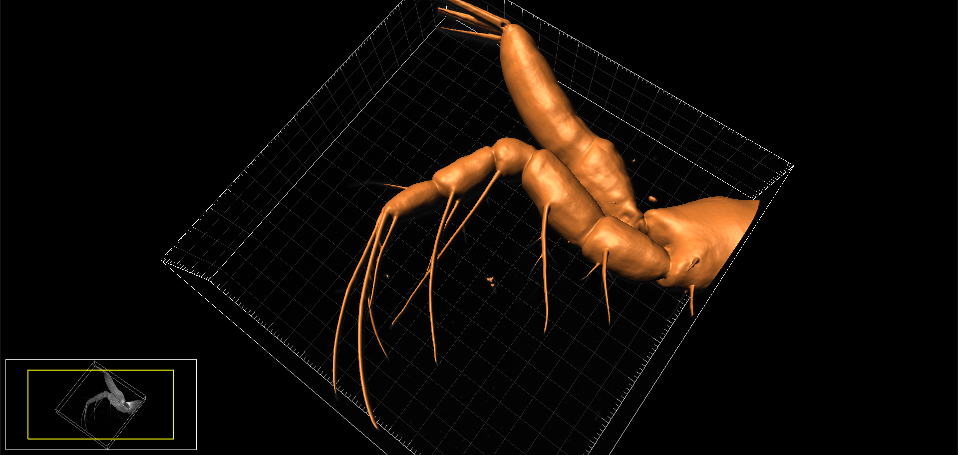
3D surface-rendered model of appendage (endopod of maxilliped) of Chinese mitten crab larva. Image produced on Zeiss LSM880 confocal and reconstructed using Bitplane Imaris.
Last week we undertook some confocal microscopy for the National History Museum to help characterise the arrangement of setae on the larval appendages of the Chinese mitten crab, Eriocheir sinesis * (now published, see Kamanli et al, 2017 below). The Mitten crab, so-named because of the tufts of ‘fur’ on the adult’s claws, is officially listed as one of the World’s most invasive species. The crabs out-compete and prey on native crab species, damage fishing nets and cause significant erosion of riverbanks, thus are of considerable economic importance. They arrived in this country from China in the 1930’s via discharge of ballast water from trading ships and are now firmly established in many of Britain’s waterways. The National History Museum is investigating ways of reducing the population of Mitten crabs and this species is currently under evaluation as a potential food source in the UK (so if you can’t beat them, eat them!)
AJH
Find out more:
- National History Museum: Chinese Mitten crab research
- The Guardian: Gloves off as scientists go to war on Mitten crab
- The Independent: Will we soon be tucking into Mitten crabs fresh from the Thames
Further reading:
- Kamanli et al. (2017) A 3D imaging and visualization workflow, using confocal microscopy and advanced image processing for brachyuran rab larvae. J Micros 266: 307-323
- Klaus & Schawaroch (2006) Novel methodology utilizing confocal laser scanning microscopy for systematic analysis in arthropods (Insecta) Integ Comp Biol 46: 207-214