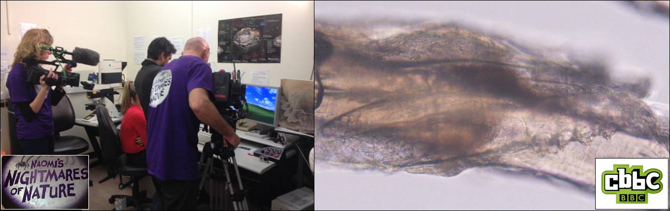
Image: BBC camera crew (left) filming the follicle mite, Demodex (right), for an episode of the children’s television programme, Naomi’s Nightmare’s of Nature.
Not so long ago we received a strange request from Dr Sarah Perkins (BIOSI): could we accommodate a BBC camera crew within the Bioimaging Facility to film an episode of the Children’s CBBC television programme, Naomi’s Nightmares of Nature? The nightmare in question, was the eyelash mite, Demodex, a commensal ectoparasite that lives within the hair follicles (Demodex follicularum) and sebaceous glands (Demodex brevis) of the face, feeding off sebum and other organic detritus. Anyway, prior to filming, we spent an anxious morning attempting to isolate live Demodex from ‘volunteers’ faces by various means, including via sellotape, with little success Fortunately, when Naomi and her production team arrived, we struck gold! A few eyelashes extracted from their cameraman, Steve, revealed a bumper load of parasites and, using DIC optics, we were able to generate some nice microscopic footage of a family of mites tucking into their evening meal!
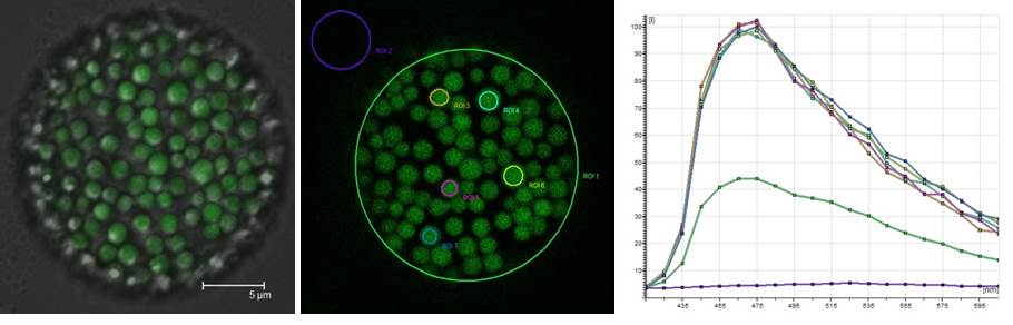
Not just pretty pictures: an earthworm coelomocyte imaged by conventional confocal microscopy (left; DIC/fluorescence image) and via spectral scanning (centre) to analyse changes in riboflavin fluorescence caused by soil pollution (right).
The Research Techniques module run by Professor Pete Kille and Dr Carsten Muller (Introduction to Environmental Toxicology) makes for a busy week within the Bioimaging Research Hub. In the practical, students learn a range of advanced analytical research techniques as they aim to identify and characterize earthworm populations that have been sampled from land polluted by heavy metal – and I’m not talking about Axel’s Rose garden here : )
Pete is an expert in ecotoxicology and much of his research centers on how invertebrate species, such as the earthworm, deal with heavy metal pollutants, e.g., lead, in their environment. As it turns out, they seem to be pretty good at tolerating a lot of the nasty stuff that passes through them, but it does leave an indelible metabolic mark – making the organisms ideal for environmental toxicological testing. And here’s where it gets interesting: previous studies of the earthworm, Eisenia fetida, have shown that heavy metals affect riboflavin (vitamin B2) biosynthesis. Now, riboflavin happens to be (1) highly autofluorescent, and (2) neatly packaged within spheroidal organelles, or chloragosomes, within a sub-population of immune cells, called coelomocytes, that are resident within the body cavity of the worm. Fortunately, earthworms can be gently persuaded to give up some of these cells for confocal microscopic analysis.
In the practical, we use confocal microscopy to image earthworm coelomocytes and, via spectral scanning, generate emission spectra of the riboflavin autofluorescence from within the chloragosomes. By comparing the autofluorescent signatures of coelomocytes from worms obtained from different sampling sites, we have asked the question: can riboflavin autofluorescence in this organism be used to assess soil pollution?
And the answer? Well, I’m not at liberty to say – the students reports aren’t in yet! (Answers on the back of a postcard to…)
Dr Amit Jathoul and Professor Jim Murray (BIOSI) have recently obtained generous funding from the Wellcome Trust to purchase a real-time, multi-spectral in vivo imaging system from Biospace Lab. The PhotonIMAGER allows bioluminescence/fluorescence imaging at a wide range of scales from cells, tissues and organs to entire complex organisms, thereby bridging the gap from single cell to whole organism imaging. Further details of the system are available through the research equipment database.
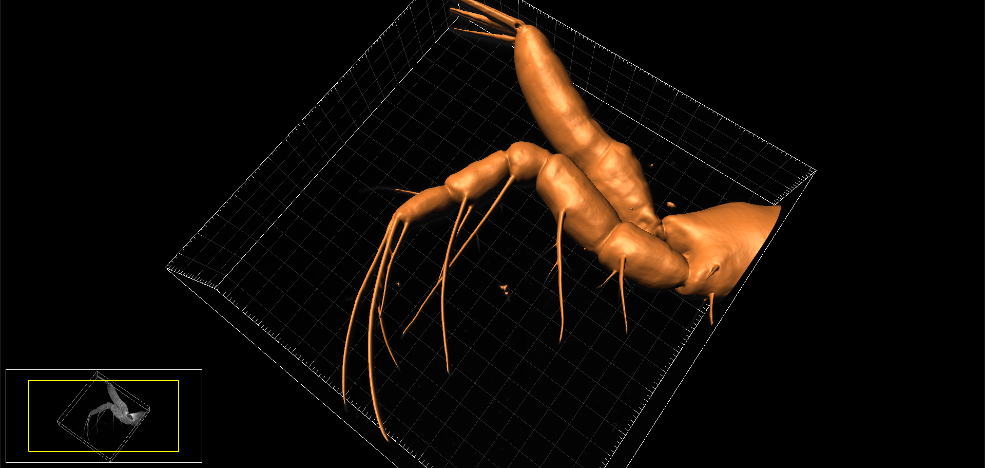
3D surface-rendered model of appendage (endopod of maxilliped) of Chinese mitten crab larva. Image produced on Zeiss LSM880 confocal and reconstructed using Bitplane Imaris.
Last week we undertook some confocal microscopy for the National History Museum to help characterise the arrangement of setae on the larval appendages of the Chinese mitten crab, Eriocheir sinesis * (now published, see Kamanli et al, 2017 below). The Mitten crab, so-named because of the tufts of ‘fur’ on the adult’s claws, is officially listed as one of the World’s most invasive species. The crabs out-compete and prey on native crab species, damage fishing nets and cause significant erosion of riverbanks, thus are of considerable economic importance. They arrived in this country from China in the 1930’s via discharge of ballast water from trading ships and are now firmly established in many of Britain’s waterways. The National History Museum is investigating ways of reducing the population of Mitten crabs and this species is currently under evaluation as a potential food source in the UK (so if you can’t beat them, eat them!)
What microscopy/bioimaging equipment do you consider essential to your research? Please take the 5 minute survey. Your response will help inform our long-term strategy for bioimaging.
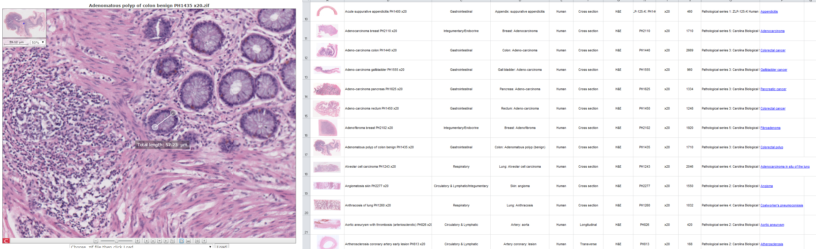
Image: The virtual histology slide box and viewer – a resource that holds fantastic potential for BIOSI teaching, research and public engagement.
The Bioimaging Hub has recently completed work in digitising the School’s extensive histopathology slide repository. Over 400 histological sections, encompassing both normal and pathological tissues, were painstakingly scanned and digitised in high resolution using the facilities Objective Imaging Surveyor slide scanning system. The datasets, totalling 4TB, have been converted into the Zoomify .ziff image file format to enable easy and rapid on-line browsing, zooming and navigation (similar to that of Google Earth) and calibrated to allow feature measurement. The image files have been linked, via thumbnails, to a database that captures all relevant metadata for each histological section (filename, tissue type, organ system, species, section plane, histological stain, section ID, supplier, objective magnification etc) to facilitate easy sorting and data retrieval. The database is currently set up on a basic Linux server within the facility; however, to cope with concurrent file access by large numbers of up to 150 students, it will require a permanent home on a dedicated server within the School. With further development, the resource promises to have fantastic potential for teaching, research and public engagement within BIOSI. Thanks to all concerned who have taken the project this far…
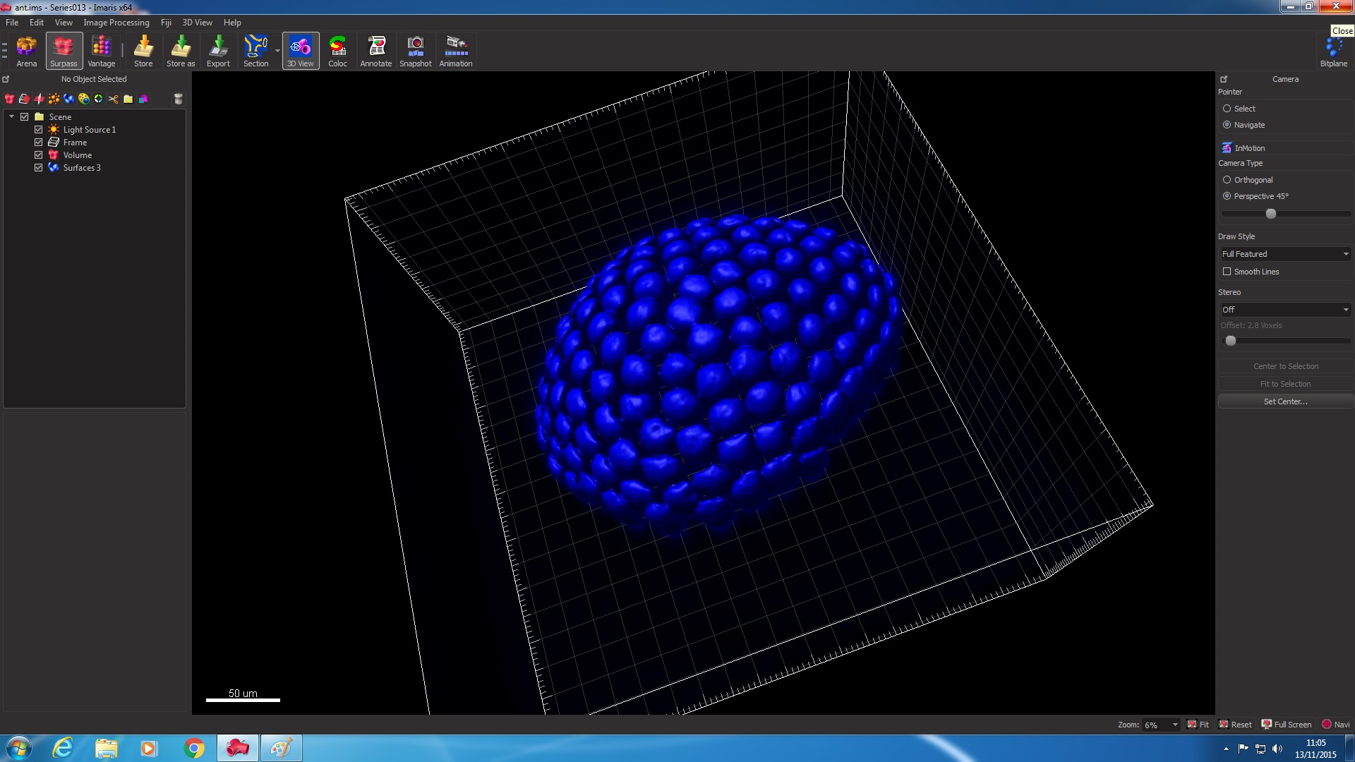
Image: Screenshot of the new Imaris software showing a surface-rendered model of an insect’s compound eye.
The Bioimaging Research Hub has recently purchased Bitplane Imaris software for Cell Biologists, together with a high-end PC workstation, via generous funding from the Research Infrastructure Fund (lead applicant: Dr Walter Dewitte). The software provides advanced processing options for confocal and multi-photon 3D/4D datasets and includes the following modules: Measurement Pro, Imaris Track, Imaris Cell, Imaris Coloc, Imaris XT and Imaris Vantage. Further details of the system are available via our equipment database.
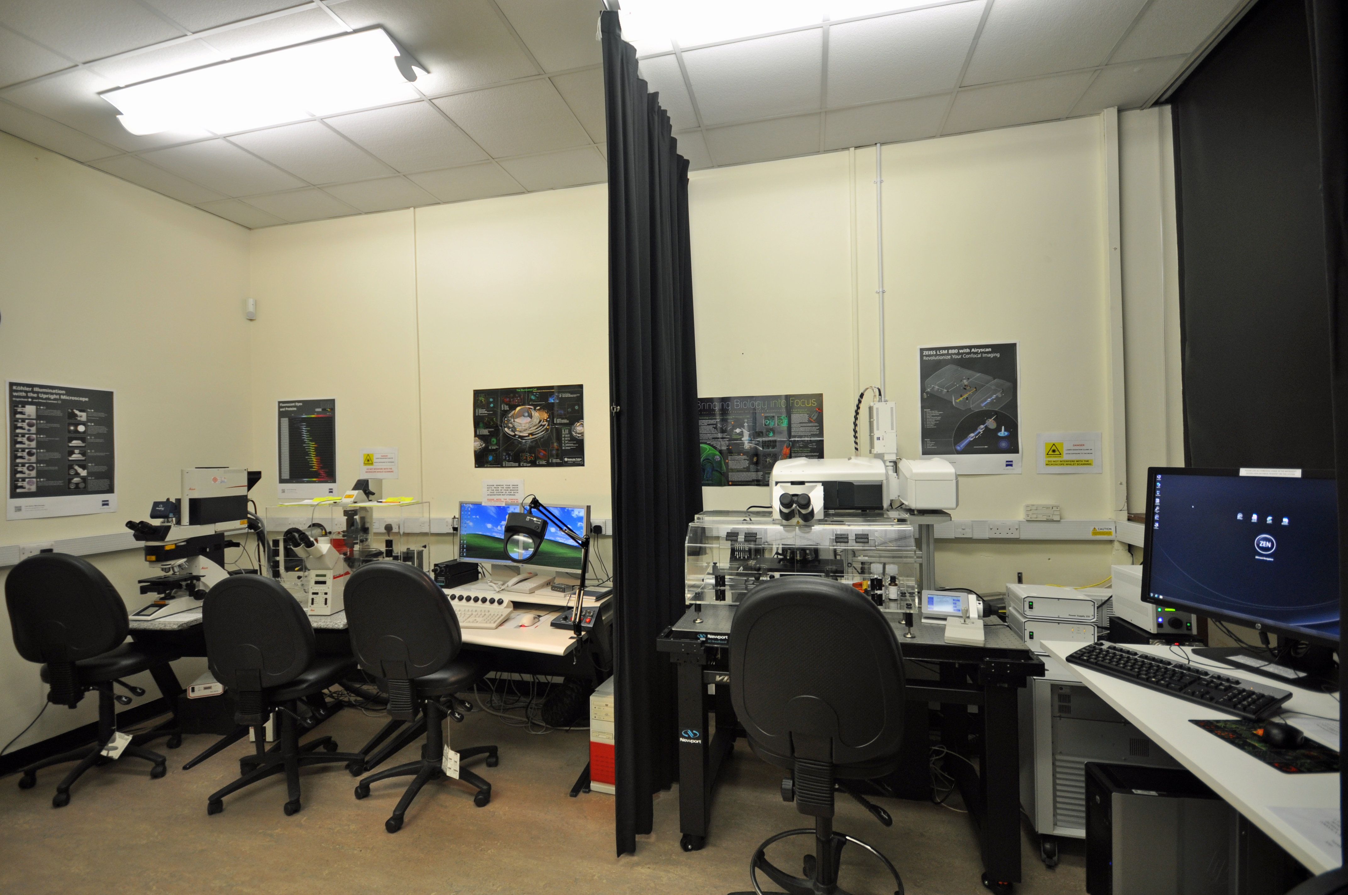
Image: The old and the new: The new Zeiss LSM 880 Airyscan confocal system (right) adjacent to the old Leica TCS SP2 confocal (left) (BIOSI 2; E/0.03)
The BIOSI Bioimaging Research Hub has recently expanded its imaging toolbox with a new, state-of-the-art confocal microscope system, that was purchased through generous funding by the Research Infrastructure Fund (Lead Applicant: Dr Walter Dewitte). The system, a top-of-the-range Zeiss LSM 880 upright confocal microscope, features the advanced Airyscan super-resolution detection module which provides a 1.7x gain in resolution in all three dimensions compared to conventional confocal optics. The system also supports advanced fluorescence techniques including FCS (fluorescence correlation microscopy) and FLIM (fluorescence lifetime imaging (FLIM) – the FLIM module will be installed during the first week of December. Further details here.
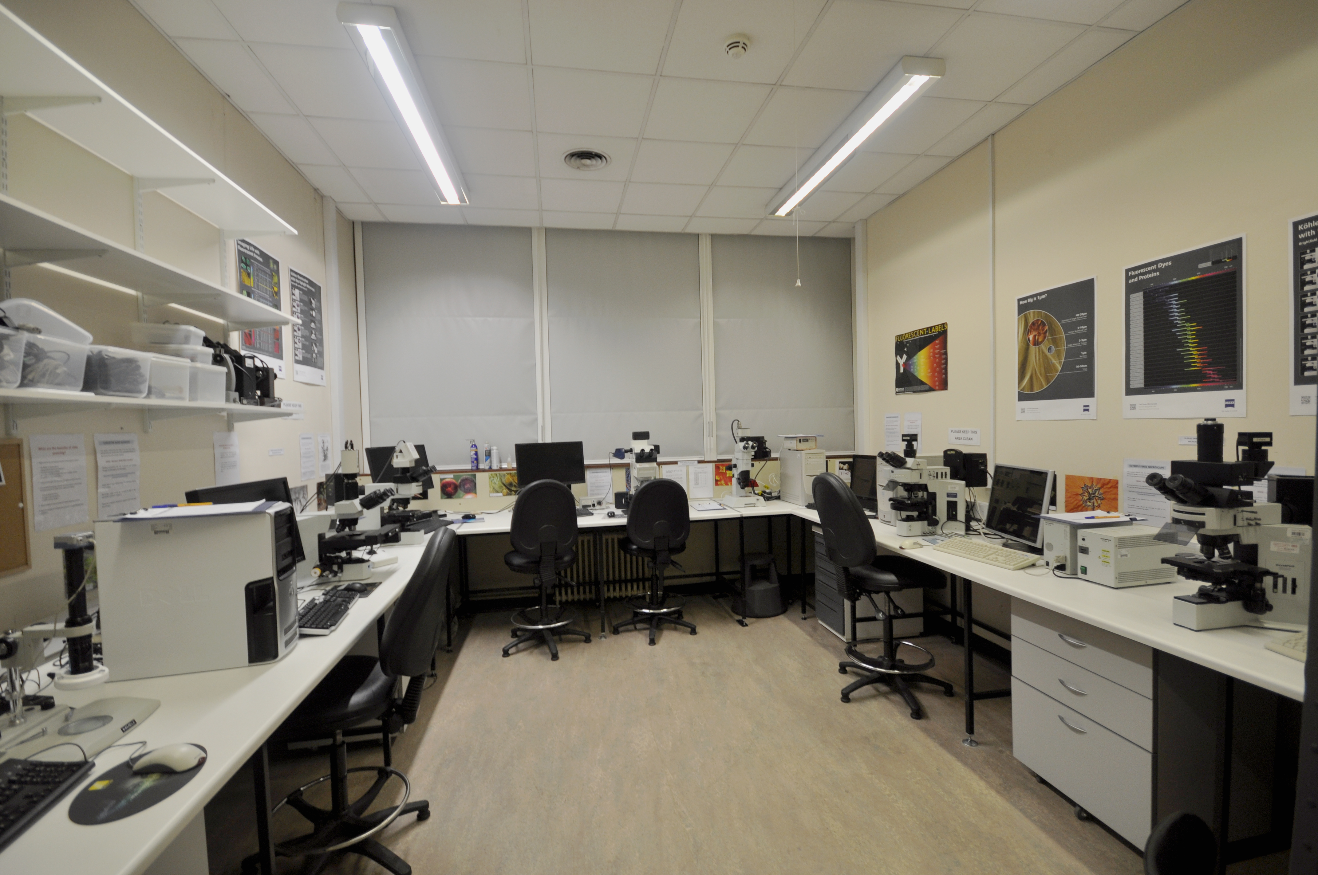
Image: The newly-refurbished microscopy suite (BIOSI 2; E/0.04).
The old SEM room (BIOSI 2; E/0.04) has now been refurbished as a dedicated widefield microscopy suite via School support. The refurbishments include new electrical and internet connections, benching, secondary glazing and black-out blinds. The suite hosts a broad range of modern transmitted light and epifluoresence systems including (from left to right):
All of the systems are available for use with or without technical support and are suitable for a wide variety of research purposes, including student projects.
This is a short post to outline what you will find in each of the sections within this blog.
Features contains information on recent events within the Bioimaging facility (e.g. new equipment, interesting research applications etc). Posts will be labelled either NEWS for general news items, IN-FOCUS for particular highlights of applications within the Unit or EQUIPMENT for new pieces of equipment in the unit.
Resources highlights some of the microscopical imaging techniques that are currently available and how these may benefit your research.
Gallery showcases a selection of the microscopical images produced within the facility.
Publications provides a list of all known publications (with linkouts) arising from research conducted within the facility.
FAQs contains answers to Frequently Asked Questions, fluorochrome data and other technical miscellany.
We hope that you find the information contained within this blog useful, or at least interesting, and look forward to your comments,