Author Archives: Anthony Hayes

IN-FOCUS: Bigging it up: 3D printing to change the shape of microscopy.

3d pollen

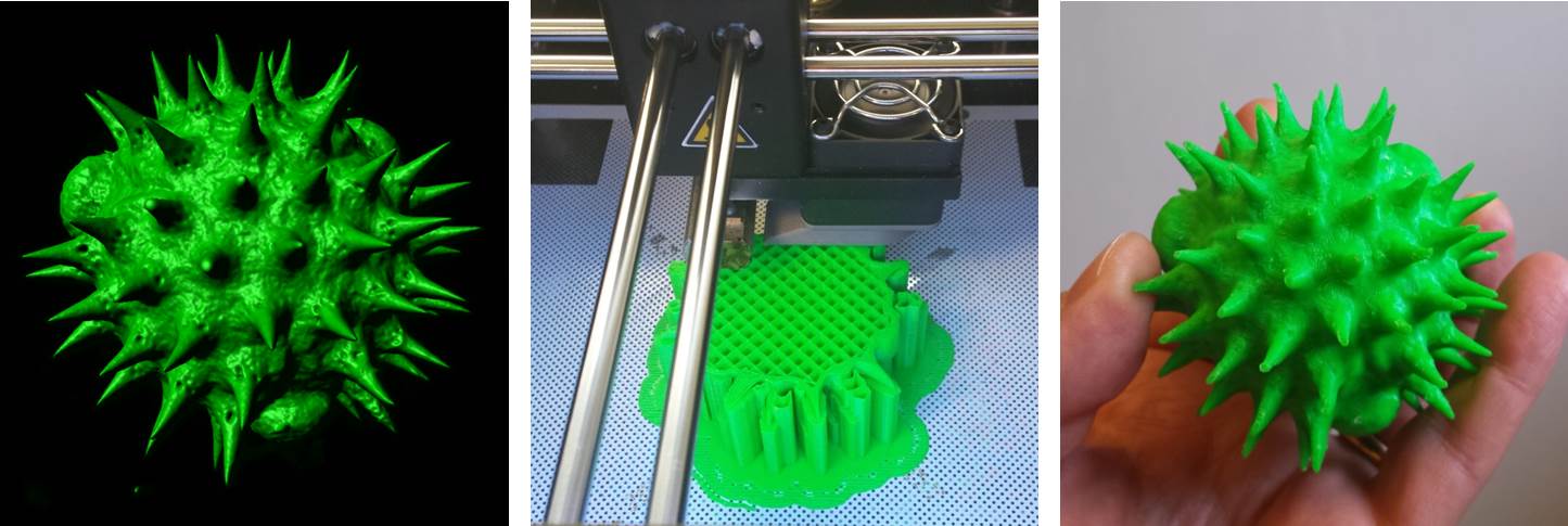
Virtual to reality: a surface-rendered digital image of a single pollen grain generated by confocal microscopy (left) is 3D printed into a 2000x scale replica model (centre & right).

Imagine being able to generate a highly accurate, solid scale replica of the sample that you are visualising down the microscope; a perfectly-rendered pollen grain, or blood cell, or microscopic organism, but big enough to hold and examine in your hand.  It would allow much better 3D conceptualisation of the sample, particularly for blind or visually-impaired individuals, and would have enormous utility in teaching and in engagement activities, and what researcher wouldn’t want a tangible, physical embodiment of their research to help explain their work (and impress their colleagues) at scientific meetings? Sounds like the stuff of science fiction doesn’t it? Well, not any more. Thanks to 3D printing technology (and the help of Dr Simon Scofield‘s lab) we have started taking volume datasets from the confocal microscope out of the virtual world and making them a reality. If you would be interested in generating a highly accurate scale model of your favourite biological sample (or would simply like to handle a giant pollen grain!) then please feel free to get in touch.

AJH

 Further reading:

IN-FOCUS: Microscopy on the move. A round-up of the best microscopy apps for mobile devices.

mobile microscope

Here’s a quick round up of some useful imaging applications for portable Android and Apple devices.

  • Molecular Probes 3D Cell App. Learn about the cell and all its structures in 3D on Apple portable devices. Enjoy the ability to rotate the cell 360 degrees and zoom in on any cell structure.

If you wish to use your smartphone camera as a rudimentary digital magnifier just search ‘microscope’ in either the Google Play or iTunes App stores – there’s loads to choose from. Instructions available here showing how to build a perspex support stage with transmitted light illumination for your smartphone. If you want anything more sophisticated, take a look at this.

AJH

IN-FOCUS: Microscopy and Analysis Journal: A Useful Resource for Microscopists.

I can see why Microscopy and Analysis is the leading international journal for microscopists – it’s  chock-full of interesting articles, features and news on all things related to microscopy and imaging. More to the point, it’s free to individuals who purchase, specify or approve microscopical, analytical and or/imaging equipment at their place of work. The journal is published six times per year, in January, March, May, July, September, and November. There are also several supplements published periodically, which include publications devoted to special events, trade shows and specific areas of microscopy and imaging. The journal is available in print,  or can be viewed online in an interactive format, or via a downloadable app. We also have lots and lots of back issues available within the Bioimaging Unit, which you are welcome to peruse on your next visit!

AJH

CORE EQUIPMENT: PicoQuant FLIM upgrade for the Zeiss LSM880 Airyscan Confocal Microscope.

FLIM demo

Image: Dr Pete Watson (left) gets to grips with the new Picoquant FLIM module. Dr Volker Buschmann of PicoQuant (right) provides expert advice.

Our new Zeiss LSM880 Airyscan confocal system has now been upgraded with the PicoQuant FLIM module for Fluorescence Lifetime IMaging. This module provides an additional two lasers (picosecond pulsed 440nm and 485nm diodes) and utilizes the Zeiss BiG.2 GaAsP detector to allow time-correlated single photon counting (TCSPC). The FLIM module is run through Picoquant Symphotime software, which integrates seamlessly with the Zeiss Zen Black confocal software.

AJH

Find out more:

NEWS: Science, Technology, Engineering & Mathematics (STEM) Conference, 2015.

STEM conference 2015

The STEM conference 2015 which featured interactive exhibitions and demonstrations by the Bioimaging Unit. Left: Six form students get to grips with 3D imaging. Right: Demonstration of the virtual histology slide  box developed by the Bioimaging Unit.

The Bioimaging facility plays a significant role in public engagement and student recruitment activities within the School of Biosciences. This summer (June 19th, 2015) the Bioimaging Facility hosted large groups of students from St David’s Catholic sixth form college as part of the Universities Science, Technology, Engineering & Mathematics (STEM) annual conference. The event, organised by Dr Fiona Wyllie, was well-attended (with over 400 students) who engaged fully in the interactive exhibitions and demonstrations, which included confocal microscopy, macro-imaging and slide scanning microscopy. As part of the slide scanning demonstration, students were able to trial a virtual histology slide box – an online digital repository of over 400 high resolution histological images – that has been developed in-house by the Bioimaging Facility for School teaching purposes. 

AJH

Find out more:


IN-FOCUS: Imaging on a Budget? A Round-up of the Best Free Imaging Software on the Web.

Grant failed to make it past triage? Departmental account looking decidedly bare? Fear not dear reader, we have trawled the net to come up with a list of the best free imaging software out there…

The following links are to downloads of free software for image acquisition, processing and multi-dimensional analysis. Hardware requirements, application notes and user instructions are all available through the individual websites. Please note that some of the downloads will require site registration.

BioImageXD    Open source software for analysing, processing and visualising multi-dimensional microscopy images.

Cell Profiler    Versatile 2D processing platform for high throughput screening applications.

Confocal Assistant    Software for 3D processing and analysis of confocal images.

Drishti    Advanced software for 3D rendering of volumetric datasets.

FluoRender    Interactive 3D rendering tool for confocal microscopy designed specifically for neurobiologists.

Icy  open community platform for bioimage informatics. Broad selection of plugins and protocols.

ImageJ    Multi-format (Java-based) open source software package for data acquisition, analysis and processing. Extensive functionality conferred via a wide selection of downloadable plugins.

LAS-AF Lite    ‘Lite’ version of the Leica application suite which allows basic processing and analysis of  image data obtained from advanced Leica widefield and confocal systems.

LCS Lite    ‘Lite’ version of the Leica confocal software that allows basic processing and analysis of Leica SP2 confocal image files.

Micro-Manager    Open source software for control of automated microscopes which runs as a plugin to Image J.

Open Microscopy Environment (OMERO)    Client server software for visualisation, management and analysis of biological images.

DeconvolutionLab    Software for  deconvolution of 2D or 3D microscopic images which runs as a plugin to Image J.

V3D    Powerful open source software for visualisation and segmentation of large 3D datasets.

View5D    Software for analysis and processing of multi-dimensional volumetric datasets which runs as a plugin to Image J.

VisBio    Open source software for visualisation and analysis of multidimensional image data. Interfaces with Image J and OMERO.

Voxx    Voxel-based rendering software for 3D analysis of confocal and multi-photon datasets.

LSM image browser    Imaging software for Zeiss LSM 5 series confocals.

ZEN Lite    ‘Lite’ version of the Zeiss Efficient Navigation  (ZEN) software that allows basic processing and analysis of image data from advanced Zeiss light microscopical systems.

AJH

IN-FOCUS: De-boning the Zebrafish: unpicking skeletogenesis under the microscope.

Confocal reconstructions of the head, thorax and tail regions of the Zebrafish (Danio rerio)

Confocal reconstructions of the head, thorax and tail regions of the Zebrafish (Danio rerio)

The Zebrafish (Danio rerio) is, in many ways, the perfect model for microscopists. Not only does it share 70% genetic homology with man, but its larvae are born in large, transparent broods all year round and develop extremely quickly (a single cell develops into something resembling a fish within 24 hours!)  This means that developmental events can be visualised in vivo in real-time down the microscope. On top of this, their genome has been sequenced and it is easily amenable to molecular manipulation- again, these manipulations can be followed closely under the microscope lens.

Over the last few years we have been collaborating with Dr Chrissy Hammond at Bristol University, a fish biologist who shares an interest in skeletal development and disease. In our joint studies, we have used a variety of imaging techniques (brightfield, DIC, polarising, epifluorescence, confocal, TEM, radiography and microCT) to investigate skeletal development, growth and ageing in  this animal model.

One of the many interesting findings from our studies is that ageing fish undergo degenerative changes to their spine that resemble osteoarthritis (for example, spinal curvature, osteophyte formation, and connective tissue degeneration). This opens up the possibility  that they could be used to experimentally model aspects of the human disease. So it’s not just fishy tails!

AJH

Find out more:
Further Reading:

IN-FOCUS: Gumming up the works: oral biofilms under the microscope.

Image showing yeast cells colonising a tissue-engineered oral epithelium

Yeast cells (Candida albicans; red) colonise and invade a tissue-engineered oral epithelium (cell nuclei; blue; cytoplasm, green)

The BIOSI Bioimaging Facility has worked closely with Professor David Williams at the Dental School in Cardiff for a number of years. David is an expert in oral microbiology, specialising in microbial biofilms (e.g. dental plaque) and mycoses such as oral candidiases (thrush). Over this time, we have been involved in a number of collaborative studies where we have used confocal microscopy and various fluorescent labelling techniques to investigate the formation, 3D organization and microbial community structure of biofilms grown on tissue engineered oral epithelium, endotracheal tubes and substrates such as dental acrylic and titanium. The research has also evaluated the effect of various anti-microbial and anti-fungal compounds and commercial mouth rinses on biofilm development using fluorescent viability stains. The studies have extended our understanding of how oral biofilms develop and in how they respond to therapeutic intervention, and have resulted in a number of publications (see below) as well as a book cover for a leading text on the subject of Oral Microbiology. It’s a fantastic application of confocal microscopy to a biological problem and, from an imaging perspective, its been something for us to really get our teeth into!

AJH

Find out more:
Further reading:

IN-FOCUS: Shedding light on new fluors: development of novel, metal-based probes for bioimaging.

Dr Simon Pope and his research group in CHEMY have been collaborating with the BIOSI Bioimaging Unit for over 8 years. Their research is focussed upon the development of new, metal-based, fluorescent probes for cell imaging applications, and forms part of a larger study on the use of metal complexes, which include rhenium and gold, as multi-modal imaging agents with therapeutic potential. In these studies, we have performed confocal imaging to (1) assess the cytotoxicity of the new probes, (2) evaluate their cellular uptake and determine their cytoplasmic localisation, and (3) characterise their fluorescent emissions via spectral (wavelength or lambda) scanning. The collaboration has yielded a number of high impact publications (see below) as well as a journal cover! With the improved potential of the new super-resolution confocal system we anticipate a lot more to come.

AJH

Find out more:

NEWS: Naomi’s nightmares of nature.

Naomi

Image: BBC camera crew (left) filming the follicle mite, Demodex (right), for an episode of the children’s television programme, Naomi’s Nightmare’s of Nature.

Not so long ago we received a strange request from Dr Sarah Perkins (BIOSI): could we accommodate a BBC camera crew within the Bioimaging Facility to film an episode of the Children’s CBBC television programme, Naomi’s Nightmares of Nature? The nightmare in question, was the eyelash mite,  Demodex, a commensal ectoparasite that lives within the hair follicles (Demodex follicularum) and sebaceous glands (Demodex brevis) of the face, feeding off sebum and other organic detritus. Anyway, prior to filming, we spent an anxious morning attempting to isolate live Demodex from ‘volunteers’ faces by various means,  including via sellotape, with little success Fortunately, when Naomi and her production team arrived, we struck gold! A few eyelashes extracted from their cameraman, Steve, revealed a bumper load of parasites and, using DIC optics, we were able to generate some nice microscopic footage of a family of mites tucking into their evening meal!

AJH

Find out more: