Category Archives: news

IN-FOCUS: Development of a 3D printed pollen reference collection.

pollen montage 1
pollen montage 2

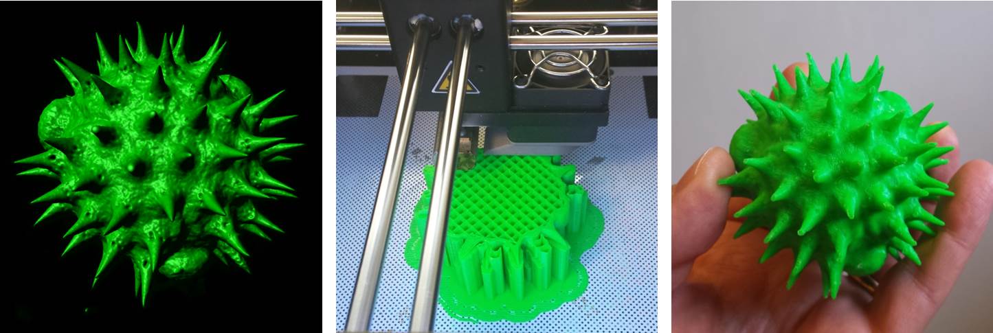
Above: surface-rendered confocal reconstructions of pollen samples (left) and their corresponding 3D printed models (right).

Isn’t the World Wide Web a wonderful thing? Not so long ago I wrote a short blog explaining how we had developed methodology to convert volume datasets from the confocal microscope into 3D printed models – perfect solid scale replicas of samples the size of a pollen grain etc. Well, shortly afterwards I received an email from someone who had not only read the blog but, serendipitously, wanted to do this very thing! What is more, she was located not a million miles away: in fact, little more than 400 yards down the road from us, working as a researcher within Cardiff University’s School of History, Archeology & Religion. Please excuse the pun, but it really is a small world!

Rhiannon Philp is an archaeologist – or palynologist to be precise – someone who studies ancient pollen grains and spores found at archaeological sites. Pollen extracted from archeological digs can be used for radiocarbon dating and for studying past climates and environments by identifying plants growing at the time. Rhiannon is using this information to develop an understanding of prehistoric sea level changes in South Wales as part of the Changing Tides Project.

Rhiannon asked if we could generate a reference collection of 3D pollen prints that could be used for teaching and outreach activities as part of a new Archaeology engagement project called Footprints In Time. Indeed, some of her pollen samples were from sites containing both human and animal footprints made over 5000 years ago!

You can see some of our results above: on the left are the surface-rendered confocal volume reconstructions and, on the right, their corresponding 3D printed facsimiles – courtesy of the BIOSI 3D printing facility.

If you’re at the National Eisteddfod in Abergavenny this week (29th July – 6th August), then please pop by to see Rhiannon’s stall within the Cardiff University tent – all of the models will be on display there, together with a lot more.  Any further interest, then please get in touch.

AJH

 Further reading:

IN-FOCUS: Bigging it up: 3D printing to change the shape of microscopy.

3d pollen

Virtual to reality: a surface-rendered digital image of a single pollen grain generated by confocal microscopy (left) is 3D printed into a 2000x scale replica model (centre & right).

Imagine being able to generate a highly accurate, solid scale replica of the sample that you are visualising down the microscope; a perfectly-rendered pollen grain, or blood cell, or microscopic organism, but big enough to hold and examine in your hand.  It would allow much better 3D conceptualisation of the sample, particularly for blind or visually-impaired individuals, and would have enormous utility in teaching and in engagement activities, and what researcher wouldn’t want a tangible, physical embodiment of their research to help explain their work (and impress their colleagues) at scientific meetings? Sounds like the stuff of science fiction doesn’t it? Well, not any more. Thanks to 3D printing technology (and the help of Dr Simon Scofield‘s lab) we have started taking volume datasets from the confocal microscope out of the virtual world and making them a reality. If you would be interested in generating a highly accurate scale model of your favourite biological sample (or would simply like to handle a giant pollen grain!) then please feel free to get in touch.

AJH

 Further reading:

NEWS: Science, Technology, Engineering & Mathematics (STEM) Conference, 2015.

STEM conference 2015

The STEM conference 2015 which featured interactive exhibitions and demonstrations by the Bioimaging Unit. Left: Six form students get to grips with 3D imaging. Right: Demonstration of the virtual histology slide  box developed by the Bioimaging Unit.

The Bioimaging facility plays a significant role in public engagement and student recruitment activities within the School of Biosciences. This summer (June 19th, 2015) the Bioimaging Facility hosted large groups of students from St David’s Catholic sixth form college as part of the Universities Science, Technology, Engineering & Mathematics (STEM) annual conference. The event, organised by Dr Fiona Wyllie, was well-attended (with over 400 students) who engaged fully in the interactive exhibitions and demonstrations, which included confocal microscopy, macro-imaging and slide scanning microscopy. As part of the slide scanning demonstration, students were able to trial a virtual histology slide box – an online digital repository of over 400 high resolution histological images – that has been developed in-house by the Bioimaging Facility for School teaching purposes. 

AJH

Find out more:


NEWS: Naomi’s nightmares of nature.

Naomi

Image: BBC camera crew (left) filming the follicle mite, Demodex (right), for an episode of the children’s television programme, Naomi’s Nightmare’s of Nature.

Not so long ago we received a strange request from Dr Sarah Perkins (BIOSI): could we accommodate a BBC camera crew within the Bioimaging Facility to film an episode of the Children’s CBBC television programme, Naomi’s Nightmares of Nature? The nightmare in question, was the eyelash mite,  Demodex, a commensal ectoparasite that lives within the hair follicles (Demodex follicularum) and sebaceous glands (Demodex brevis) of the face, feeding off sebum and other organic detritus. Anyway, prior to filming, we spent an anxious morning attempting to isolate live Demodex from ‘volunteers’ faces by various means,  including via sellotape, with little success Fortunately, when Naomi and her production team arrived, we struck gold! A few eyelashes extracted from their cameraman, Steve, revealed a bumper load of parasites and, using DIC optics, we were able to generate some nice microscopic footage of a family of mites tucking into their evening meal!

AJH

Find out more:

IN FOCUS: Development of a Virtual Histology Slide Box.

histology database & viewer

Image: The virtual histology slide box and viewer – a resource that holds fantastic potential for BIOSI teaching, research and public engagement.

The Bioimaging Hub has recently completed work in digitising the School’s extensive histopathology slide repository. Over 400 histological sections, encompassing both normal and pathological tissues, were painstakingly scanned and digitised in high resolution using the facilities Objective Imaging Surveyor slide scanning system.  The datasets, totalling 4TB, have been converted into the Zoomify .ziff image file format to enable easy and rapid on-line browsing, zooming and navigation (similar to that of Google Earth) and calibrated to allow feature measurement. The image files have been linked, via thumbnails, to a database that captures all relevant metadata for each histological section (filename, tissue type, organ system, species, section plane, histological stain, section ID, supplier, objective magnification etc) to facilitate easy sorting and data retrieval. The database is currently set up on a basic Linux server within the facility; however, to cope with concurrent file access by large numbers of up to 150 students, it will require a permanent home  on a dedicated server within the School. With further development, the resource promises to have fantastic potential for teaching, research and public engagement within BIOSI. Thanks to all concerned who have taken the project this far…

AJH

Find out more:


NEWS: Newly-refurbished microscopy suite.

DSC_0456

Image: The newly-refurbished microscopy suite (BIOSI 2; E/0.04).

The old SEM room (BIOSI 2; E/0.04) has now been refurbished as a dedicated widefield microscopy suite via School support. The refurbishments include new electrical and internet connections, benching, secondary glazing and black-out blinds. The suite hosts a broad range of modern transmitted light and epifluoresence systems including (from left to right):

All of the systems are available for use with or without technical support and are suitable for a wide variety of research purposes, including student projects.