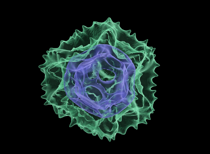
Above: Transparent 3D rendering of a dandelion (Taraxacum officinale) pollen grain. Surface exine displayed in green, inner intine structure in blue. Image produced by Dr Anthony Hayes, Bioimaging Hub, Cardiff School of Biosciences.
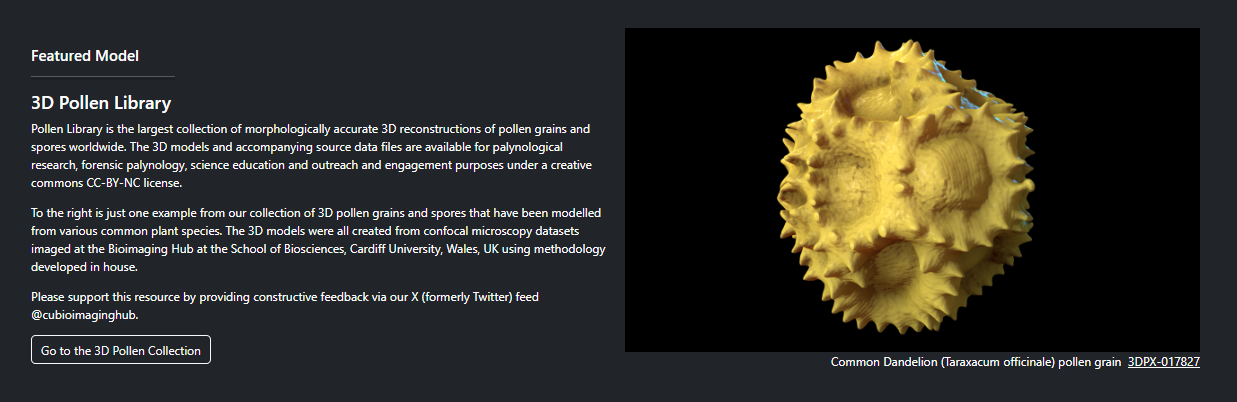
Over the last 12 months I’ve been working closely with biovisualisation specialist Dr Kristen Brown at NIH3D to curate our 3D pollen model resources into a purpose-built 3D collection: the ‘3D Pollen Library’. This collection is now featured on the NIH3D homepage and, at the time of writing, represents the largest collection of 3D pollen grain modelsworldwide. To date, it contains over a hundred entries together with taxonomic metadata and links to other well-established online pollen resources – a significant achievement considering its humble beginnings (you can read more about the background work leading up to it here.)
Enormous thanks must go to Kristen and her team who have been incredibly helpful throughout in accommodating my (many) requests regarding the ‘look and feel’ of the curated collection. They have done an absolutely splendid job.
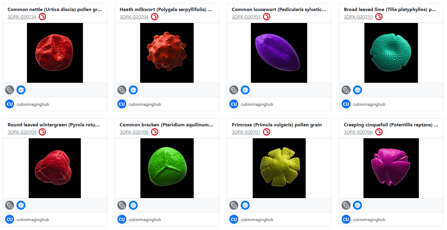
Above: A small selection of surface rendered pollen grains in the 3D Pollen Library collection. These examples were recently created in collaboration with Dr Heather Pardoe, Amgueddfa Cymru, National Museum Wales.
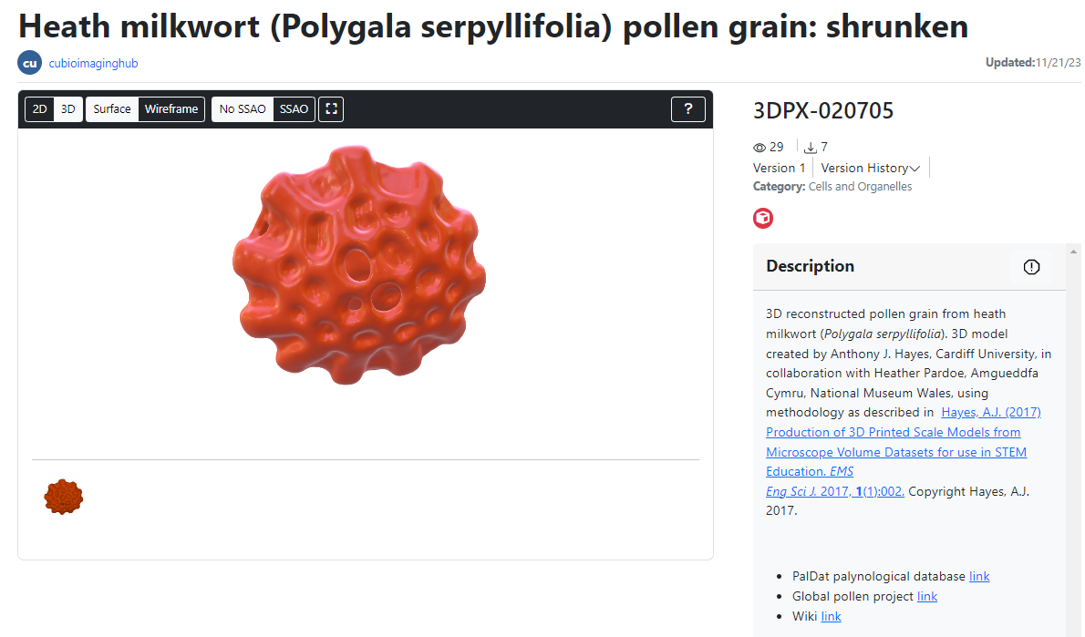
Above: the user interface for visualisation and manipulation of the 3D pollen models. The models are all viewable as surface rendered or wireframe meshes and can be downloaded in x3d, stl, glb and wrl file formats for 3D printing and AR/VR visualisation. The source confocal data is also available for download, as is the published methodology for creating the models.
In parallel with the above, I’ve established an ongoing collaboration with Dr Heather Pardoe (senior botanist and chief palynologist at Amgueddfa Cymru, National Museum Wales). This collaboration has allowed us to digitise and 3D model pollen grains and spores from selected plant species in the museum’s extensive archival pollen collection using methodology we’ve developed in-house at the Bioimaging Hub. The 3D pollen models produced via this collaboration will be added to our NIH3D library as a separate pollen sub-collection, as well as being viewable as part of the full collection. It is envisaged that these models will have significant utility as educational tools for teaching and exhibition.
Please feel free to visit the 3D Pollen Library and download some of our models (they’re all free!) We’d welcome constructive feedback with photographs via our X (twitter) feed @cubioimaginghub which will allow us to continue developing and improving the 3D resource.
Above: A selection of stage plate inserts 3D printed by the Bioimaging Research Hub – links to resources in blog article below.
Hands up if your microscope is badly in need of upgrade or repair but your budget won’t stretch that far? Maybe a new focusing knob to replace the one that just broke off in your hand, or perhaps a new stage plate adapter, reflector cube or filter holder to increase your imaging options? Perhaps a C-mount or smartphone adaptor to give one of your old microscopes a new lease of life? Or even a sample holder or chamber for a bespoke imaging application? What the heck, let’s think big eh? How about a completely new modular microscope system with tile scanning capabilities?
Way too expensive, eh?… Well, imagine for a moment that you could just click a button (or a few buttons, at least) and make it so. If you haven’t yet realised, I’m talking about 3D printing in light microscopy and the life sciences – the subject of a very interesting paper that I recently came across – see below.
It’s safe to say that 3D printing is changing the way we do things in microscopy, now permitting low-cost upgrade, repair, or customisation of microscopes like never before. There are now a huge selection of 3D printable resources available through websites such as NIH3D, Thingiverse etc that can be used to modify your microscope system or to generate scientific apparatus or labware for upstream sample processing and preparation procedures.
So, to save you trawling through the 3D printing sites in order to identify the most useful designs to meet your histology and imaging needs we’ve done it for you and have curated a list of 3D printable resources which we hope you you’ll find useful (below).
3D printable resources for histology and light microscopy. Information collated by Dr Tony Hayes, Bioimaging Research Hub, School of Biosciences, Cardiff University, Wales, UK.
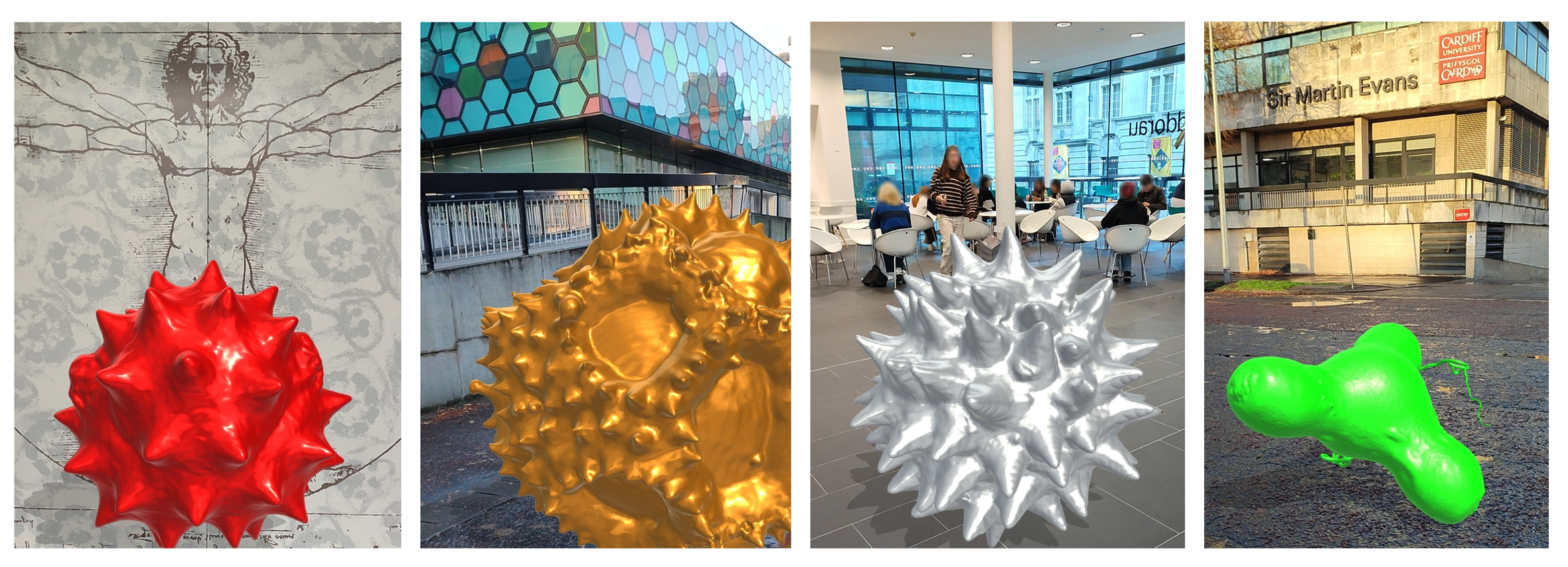
Above: Augmented reality visualisation of pollen grains 3D rendered in gigantic proportions. You wouldn’t want one of these getting stuck up your nose!
Back in 2015 we developed some novel methodology that allowed us to generate 3D printed models from confocal z-stacks of microscopic samples. At the time, we showcased the technique by making 3D prints of pollen grains from various plant species for use in science education and outreach activities, musing that a physical, tangible model would allow improved 3D conceptualization of these microscopic structures – original blog article here. The work generated a fair amount of interest and resulted in requests for 3D printed models of pollen grains from far and wide (e.g., the Smithsonian National Museum of Natural History in the US, the UK Met Office, National Botanical Garden of Walesetc). Following publication of our methodology in 2017 other researchers soon followed suit, creating their own 3D printed models from confocal datasets utilizing methodssimilar to ours. By this time, we had turned our attention to virtual reality (VR) as a tool to experience/manipulate microscopic objects, as well as larger more complex biological models in virtual space – something we saw as a logical evolution of the 3D learning experience. You can read more about this in a separate blog and see the progress we made in developing the resource via a short video on our YouTube site.
Fast-forward to present, a global pandemic, accelerating environmental change, and a cost-of-living crisis – the economic fallout from COVID, the folly of Brexit, a war in Ukraine, and gross government incompetence. To borrow a quote from a certain Vladimir Ilyich Ulyanov, there are decades where nothing happens, and there are weeks where decades happen. Well, quite!
But what of the 3D pollen models? Well, for one, coronaphobia has meant that people are less inclined these days to be handling and passing around shared resources such as plastic pollen models or VR headsets at risk of spreading/contracting germs. Moreover, plastic is increasingly being viewed negatively due to the environmental damage caused by plastics and ‘frivolous’ 3D printing could be seen as increasing the existing plastic problem. Lastly, with the cost-of-living crisis/looming recession, it would seem that no one can afford to do anything other than feed themselves and try to keep warm these days, let alone commission 3D pollen prints from the Bioimaging Hub – particularly since there’s now a growing number of pollen models available through 3D printing sites such as Sketchfab etc. And so, with these concerns in mind, we’ve donned our thinking caps and come up with a plan to address the prevailing zeitgeist.
Firstly, we’ve decided to make our entire repository of 3D pollen models publicly available, free of charge, under a Creative Commons CC-NY-NC licence via the newly developed NIH3D website (formerly the NIH3D print exchange) which is a leading community-driven portal for sharing and downloading bioscientific content for 3D printing and interactive 3D visualization. Under the terms of our licence, the models can be used, shared, or modified for non-commercial purposes, as long as the creator(s) are properly credited. To date, we’ve made available 3D models of seventy distinct species of pollen grains and spores through the Bioimaging Hub’s new NIH 3D profile page which have now been curated into a special collection, the 3D Pollen Library We have also included all source data (confocal z-stack files in Carl Zeiss Image .czi file format). To the best of our knowledge, this represents the largest single collection of 3D pollen files available online and the plan is to add more models plus supporting data in future. For cross-referencing, relevant links have been included to major palynological databases, PalDat and the Global Pollen project, and, of course, Wikipedia – the fount of all knowledge : ) We’ve also showcased a small selection of our pollen models on the Bioimaging Hub’s Sketchfab site – these can’t be downloaded directly, however you can manipulate the models on screen and view them in VR – further information here.
Secondly, we’ve been experimenting with augmented reality (AR) as a tool to allow visualization and exploration of our 3D models in real-world environments. The beauty of AR, of course, is that it doesn’t require a headset, just a smartphone or tablet which are now more ubiquitous than ever. Thus, a 3D model in the relevant file format (usually .glb or .gltf) can be downloaded directly to an individual’s smartphone or tablet and, using appropriate AR software, seamlessly integrated with real time digital information from the camera for display on the touchscreen. This allows users to personally experience a real environment with generated perceptual information overlaid upon it. Within the AR environment, the models can be scaled up or down, freely moved and rotated via the touchscreen, or circumnavigated and explored both externally and internally via directional information from the device’s sensors,à la Pokemon-GO. This allows for a highly realistic and immersive interactive experience (different from the artificial environments of VR) thus facilitating 3D conceptualization of the embedded model. Furthermore, freed from the physical encumbrance of a VR headset, the user is less likely to blunder into office furniture, moving traffic etc or experience the nausea, headaches and dizziness of VR-associated cybersickness.
So how does one view our 3D pollen models in AR? Well, it’s quite straightforward really, but to make things even easier I’ve put together a set ofinstructions (below) that should get you up and running in next to no time:
Install a free AR app on your mobile device (smartphone or tablet) from the Google Play or Apple App stores that handles .glb (GL transmission format binary) files – this is the common standard file format for AR/VR visualisation. There are quite a lot of AR apps to choose from and we’ve only tested the NeoSpace AR app on android devices but this seems to work quite well.
Go to the Bioimaging Hub’s NIH 3D webpage – link here – select a 3D pollen model, and then click on the DOWNLOAD option. You will then see a list of the files that we’ve made available for download (refer to screenshot below). These include:
.glb file format for AR/VR applications (available as both zipped and unzipped files).
.stl (stereolithography, .wrl (‘worlds’ virtual reality modelling) and x3d file formats for 3D printing applications.
source confocal data in .czi (Carl Zeiss Image) file format (zipped file).
For AR viewing, download the .glb file to your mobile device (if zipped then extract the compressed file).
Open the .glb file in the AR viewer app and follow the on-screen instructions. Initially you’ll have to scan your environment with the camera on your smartphone/tablet so that the app can identify a flat surface in order to place the 3D model into your display.
If you’ve done it correctly then you should now be able to view any of our 3D pollen models in whatever context your imagination, mobile device and AR app allows. Below are some examples of the type of AR imagery that we’ve generated using a free android AR app.
Above: Video sequence captured from an android smartphone running the free AR app, NeoSpace. The 3D pollen model can be zoomed, freely moved and rotated via touchscreen and also circumnavigated and explored both externally and internally via directional information from the device’s sensors.
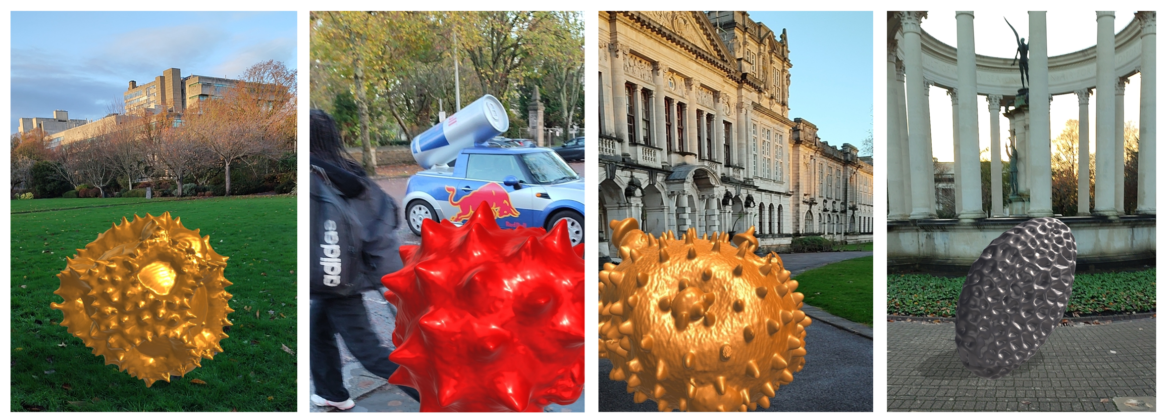
Above: Attack of the giant pollen grains! Photo-realistic AR imagery generated using an android smartphone and the NeoSpace AR app.
We’d love to get some feedback with photos or videos showing how you have used our pollen models for 3D printing, or AR/VR applications in science education and outreach. Please keep us updated via our twitter account @cubioimaginghub. Best of luck!
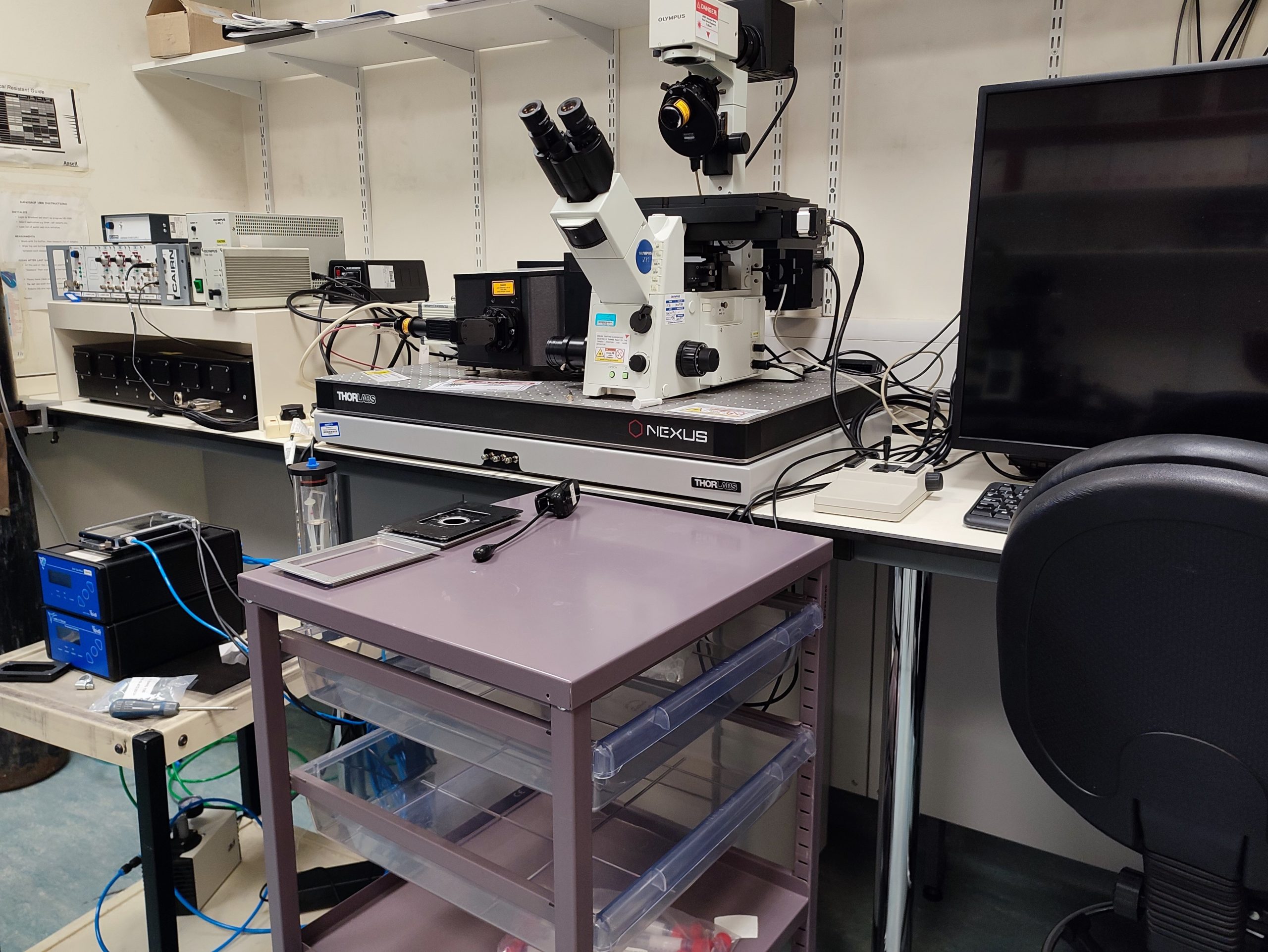
Above: Spinning disc confocal microscope set up in the new BIOSI live cell imaging spoke
A dedicated live cell imaging spoke has been set up in the Sir Martin Evans Building (BIOSI; E/3.15). The microscopy suite has inverted widefield, scanning and spinning disc confocal microscopes with full environmental control systems. Further information available through Pete Watson.
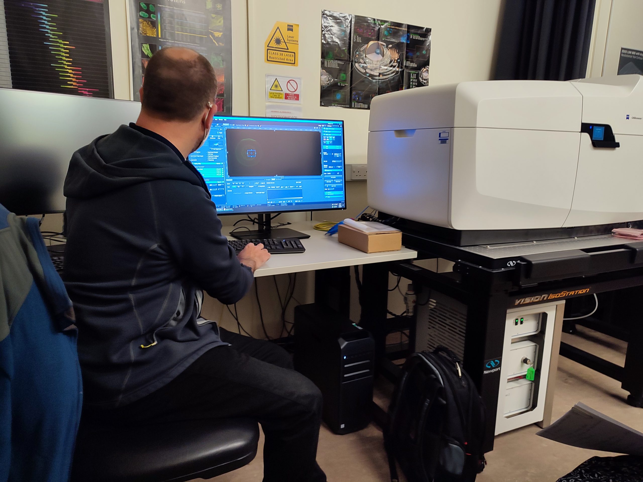
Above: Training on the Bioimaging Hub’s new Zeiss Celldiscover7 imaging system
The Bioimaging Hub has recently taken delivery of a state-of-the-art, automated live cell imaging system to replace its old Leica SP2 confocal microscope. The Celldiscoverer7 imaging system, which was purchased via the generous support of Cardiff University’s Research Infrastructure Fund (Lead Applicant: Dr Tony Hayes), has the latest Zeiss LSM 900 confocal scan head with Airyscan 2 detector technology and is capable of multi-format, high-throughput and super-resolution analysis of a wide range of samples from cell cultures to small model organisms. The system supports photomanipulation via FRAP, FRET and related techniques and is furnished with a comprehensive Zen software package that includes modules for deconvolution and machine learning, amongst other cutting edge features. Further details of the system are available through the Bioimaging Hub’s research equipment database.
A huge and heartfelt thank you to all users and support staff of the Bioimaging Hub for your strict adherence to our covid security measures over the last 12 months. It has been an extremely difficult year for all of us and we have tried to manage the situation as effectively and as safely as possible, working within the security framework provided by Cardiff University and Welsh government.
In line with the latest guidance, we are pleased to now begin relaxing some of our covid security measures and to be in a position where we can reintroduce direct hands-on support and training for our microscope systems. To facilitate this provision, it remains vitally important that users follow the new guidance protocols, as detailed below.
Users must not visit the Bioimaging Hub if they are displaying any symptoms of Covid-19; if they have been in a high risk area; or had recent contact with a covid-positive individual without confirmation of a negative test result.
All users are advised to make regular use of the Cardiff University covid screening service or to utilise rapid flow testing methodology that is now widely available through the NHS and pharmacies.
Room occupancy status and technical support/training:
Room occupancy status has now increased to two independent users/two research bubbles per microscopy suite, up to a maximum of four people per microscopy suite in total.
Direct hands-on training and support for our microscope systems will now resume under social distancing rules and with PPE including face coverings.
COVID working Regulations within the Bioimaging Research Hub:
Users should continue to use the room booking calendars and should specify which microscope system they require in the title section of the booking request (i.e., specify user name and microscope system). Booking details here.
Social distancing rules remain in place within all areas of the Bioimaging Hub.
PPE remains mandatory including the use of face coverings in all communal areas.
Microscope cleaning procedures before and after use remain mandatory.
Histology sample drop-offs and collections should continue to be arranged via email (bioimaginghub@cardiff.ac.uk).
With the dizzying pace of technological innovation and ongoing advances in microscopy and imaging, it is becoming increasingly difficult to keep abreast current developments in the field. With this in mind, we’ve rounded up some essential resources, from basic to advanced, that will keep you informed and updated.
Above: the new Olympus VS200 high throughput slide scanning system in ECSCRI
A new, high throughput slide scanning system has recently been installed in the European Cancer Stem Cell Research Institute (ECSCRI) and is available for use as a spoke of the Bioimaging Research Hub. The equipment allows automated high-throughput scanning of histological samples via a range of image modalities, including epifluorescence. Further details of the system are available through the Hub’s equipment database. All enquiries for this system should be directed towards Mr Mark Bishop.
For the time being, use of the Bioimaging Hub is going to be a very different experience for everyone.In drawing up these guidelines we’ve taken a lead from the Welsh Government and have started with a very cautious approach that we’ll constantly review and relax where possible. For now, the guidance may seem quite stringent so please bear with us while we all adapt to this new way of working.
Access is not permitted to the Bioimaging Research Hub without (i) completing a risk assessment form that should cover all planned activities within the facility and (ii) reading all relevant supporting information (see below). A template risk assessment is available through the Bioimaging Hub’s main web pages.
Before completing the risk assessment form, all users must download and read the following documents:
If you are SARS-Cov-2 positive or are displaying symptoms of COVID-19 infection (e.g. persistent coughing, elevated temperature, anosmia, sickness).
If you have been in a high-risk area or have had recent contact with confirmed SARS-CoV-2 positive individuals within the last 14 days.
If you have an underlying health condition and are concerned it will put you at greater risk of developing severe COVID-19 symptoms. N.B. essential work can be undertaken by Bioimaging Hub staff at a supported rate if necessary.
COVID Working Regulations within the Bioimaging Research Hub
All users of the facility:
MUST read the above documentation before entering the Bioimaging Research Hub.
MUST contact bioimaginghub@cardiff.ac.uk before entering the facility.You are not permitted to simply drop-in unplanned. Drop off/collection of histology samples must be arranged in advance. Researchers must knock before entering histology via E/0.08 and observe all safety directives outlined in this document. Users must complete a histology request form to specify processing preferences in advance of their visit.
MUST wash their hands upon entering and leaving the facility (hand wash station opposite office area). Multiple gel hand wash dispensers are also available within the microscopy suites.
MUST wear appropriate personal protective equipment (PPE) within the facility, including a buttoned-up lab coat and gloves (gloves are provided in all microscopy suites). Eye protection is advisable.
MUST clean the imaging equipment and immediate working area before and after use (cleaning instructions are available at each microscope station; alcohol spray and lab roll have been provided for this purpose).
MUST observe social distancing (i.e. 2 metres inter-personal space) within the Hub. The main corridor should be used by only one person at a time. While social distancing measures are in place no training is available and technical support will be provided on a remote basis via telephone (main office: 02920876611; shared office: 02922510220; histology: 02920875139).
MUST NOT enter the staff office area (0.14A) or histology suites (E/0.06-E/0.07). Drop off/collection of histology samples must be arranged in advance (use E/0.08).
MUST knock before entering any room. All microscopy suites are now single occupancy (i.e. one in, one out with at least 15 mins user-free time between bookings). The microscope booking calendars have been replaced with room booking calendars, as follows: “BIOSI – E/0.03 – Confocal/Lightsheet microscopy”, “BIOSI – E/0.04 – widefield microscopy”, “BIOSI – E/0.05 – spinning disc microscopy”. New booking instructions can be found here.
MUST set the room occupancy status (vacant/in use) on the door sliders of the microscopy suites before and after use.
Above: the new look Bioimaging hub YouTube channel.
With the University (and entire planet) in lockdown due to the ongoing covid 19 pandemic, now seems as good time as any to make you aware of the Bioimaging hub’s ‘rebooted’ YouTube channel, if you was not already aware of it.
A decade is a long time in imaging. Way back in 2009 we set up a YouTube channel to showcase the capability of our new, all-singing, all-dancing Leica SP2 confocal microscope. At the time, we uploaded a collection of short 3D animation sequences that highlighted some of our ongoing research applications. Fast forward eleven years. Whilst many of those early demo videos have been highly viewed (one over 20 thousand times) they’re starting to look rather dated, particularly when compared to the material that we’re now producing using our new confocal and lightsheet systems and 3D analysis software. As I say, a decade is a long time in imaging.
So, with the advent of spring, we thought it was high time we dusted down our YouTube channel and gave it an overhaul. As well as introducing lots of nice new image content from some of our latest 3D imaging systems, we felt that the channel would have a greater sense of purpose if we were to develop it into an educational resource for microscopy and bioimaging, with obvious relevance for remote learning (i.e., perfect in our current circumstances). Consequently, we have started to collate the most useful and relevant of YouTube’s microscopy-related content (webinars, tutorials, demonstrations etc), ranging from the basic principles of light microscopy to cutting-edge fluorescence-based nanoscopy techniques such as FLIM, so that they are all placed under one roof for your convenience : )
One of the things we hoped to provide our userbase was a series of video tutorials for the hub’s many microscope systems. Whilst there’s a great deal of useful training material on YouTube, in the main, it tends to be aimed at many of the high-end, turn-key imaging systems. Furthermore, not all microscopes are created equal, they each have their own peculiarities which reflect their intended function and most ‘evolve’ over time, through upgrades, to accomodate the vagaries of research. So, with this in mind, we have started to create our very own bespoke training videos for each of the hub’s microscope systems (example here).
The new training videos will supplement the standard operating procedures (SOPs) we have written for all of our imaging equipment and should provide an invaluable resource for user training and e-learning. As such, they will be embedded within the appropriate sections of the hub’s SOP repository (read more here). The online video content and their associated SOPs will be viewable at the click of a mouse button via desktop shortcuts on all of our microscope-associated PCs allowing easy access during instrument operation.
If you have time on your hands, then please pop over to YouTube and take a look at how our channel is developing (link here). It’s still work in progress but, as I say, it has the potential to be an extremely useful resource; not only for hub users, but anyone with a passing interest in microscopy and bioimaging. Constructive feedback is welcomed.