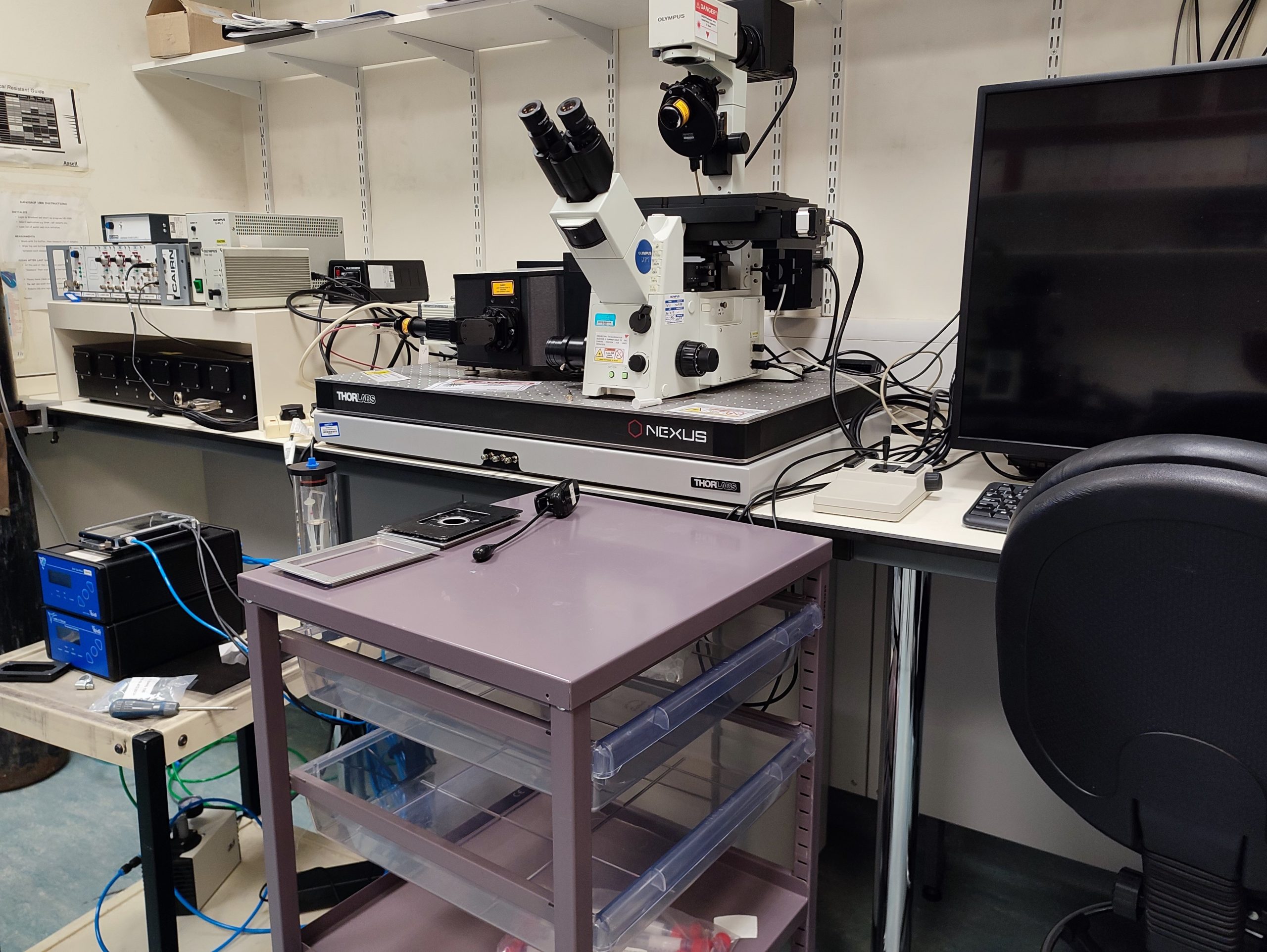
Above: Spinning disc confocal microscope set up in the new BIOSI live cell imaging spoke
A dedicated live cell imaging spoke has been set up in the Sir Martin Evans Building (BIOSI; E/3.15). The microscopy suite has inverted widefield, scanning and spinning disc confocal microscopes with full environmental control systems. Further information available through Pete Watson.
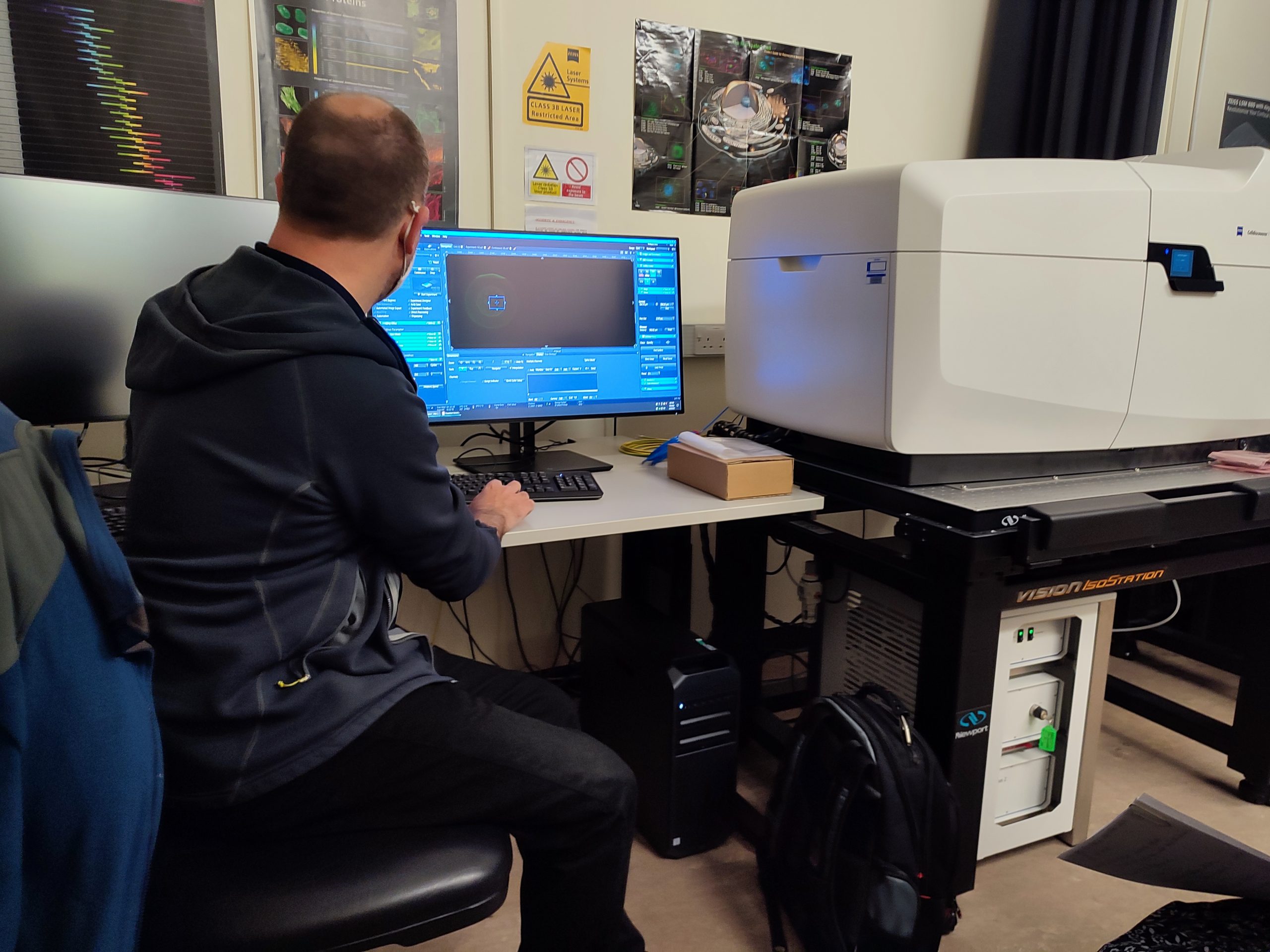
Above: Training on the Bioimaging Hub’s new Zeiss Celldiscover7 imaging system
The Bioimaging Hub has recently taken delivery of a state-of-the-art, automated live cell imaging system to replace its old Leica SP2 confocal microscope. The Celldiscoverer7 imaging system, which was purchased via the generous support of Cardiff University’s Research Infrastructure Fund (Lead Applicant: Dr Tony Hayes), has the latest Zeiss LSM 900 confocal scan head with Airyscan 2 detector technology and is capable of multi-format, high-throughput and super-resolution analysis of a wide range of samples from cell cultures to small model organisms. The system supports photomanipulation via FRAP, FRET and related techniques and is furnished with a comprehensive Zen software package that includes modules for deconvolution and machine learning, amongst other cutting edge features. Further details of the system are available through the Bioimaging Hub’s research equipment database.
A huge and heartfelt thank you to all users and support staff of the Bioimaging Hub for your strict adherence to our covid security measures over the last 12 months. It has been an extremely difficult year for all of us and we have tried to manage the situation as effectively and as safely as possible, working within the security framework provided by Cardiff University and Welsh government.
In line with the latest guidance, we are pleased to now begin relaxing some of our covid security measures and to be in a position where we can reintroduce direct hands-on support and training for our microscope systems. To facilitate this provision, it remains vitally important that users follow the new guidance protocols, as detailed below.
Users must not visit the Bioimaging Hub if they are displaying any symptoms of Covid-19; if they have been in a high risk area; or had recent contact with a covid-positive individual without confirmation of a negative test result.
All users are advised to make regular use of the Cardiff University covid screening service or to utilise rapid flow testing methodology that is now widely available through the NHS and pharmacies.
Room occupancy status and technical support/training:
Room occupancy status has now increased to two independent users/two research bubbles per microscopy suite, up to a maximum of four people per microscopy suite in total.
Direct hands-on training and support for our microscope systems will now resume under social distancing rules and with PPE including face coverings.
COVID working Regulations within the Bioimaging Research Hub:
Users should continue to use the room booking calendars and should specify which microscope system they require in the title section of the booking request (i.e., specify user name and microscope system). Booking details here.
Social distancing rules remain in place within all areas of the Bioimaging Hub.
PPE remains mandatory including the use of face coverings in all communal areas.
Microscope cleaning procedures before and after use remain mandatory.
Histology sample drop-offs and collections should continue to be arranged via email (bioimaginghub@cardiff.ac.uk).
Above: the new Olympus VS200 high throughput slide scanning system in ECSCRI
A new, high throughput slide scanning system has recently been installed in the European Cancer Stem Cell Research Institute (ECSCRI) and is available for use as a spoke of the Bioimaging Research Hub. The equipment allows automated high-throughput scanning of histological samples via a range of image modalities, including epifluorescence. Further details of the system are available through the Hub’s equipment database. All enquiries for this system should be directed towards Mr Mark Bishop.
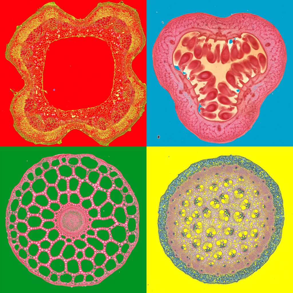
Above: Plant parts x4 . Biological pop-art on the ‘Warhols’ of the Bioimaging hub. Slide scanned images of plant tissues (various species; transverse sections) courtesy of Dr Tony Hayes, pop-art montage by Marc Isaacs.
The eagle-eyed amongst you may have noticed that the main corridor within the Bioimaging Research Hub is looking a little nattier these days. We thought it required some brightening up and so have started to adorn its walls with some nice, new A0-sized foamex prints of microscopical images that we’ve generated in-house on the Hub’s microscope systems. We’ve tried to select images that showcase the beauty of the unseen microscopic world which reflect the art in science, or ‘SciArt’ as it is known. We hope the images stimulate interest and highlight the state-of-the-art research and facilities within Cardiff School of Biosciences. No prizes for spotting the artistic influences on some of our ‘works’ : )
The Bioimaging research hub does a nice sideline 3D printing scale replicas of biological samples for use in science engagement and teaching. These can be made from the smallest of microscopic samples imaged via optical sectioning microscopy (i.e., confocal or lightsheet) or from large anatomical samples imaged via 3D photogrammetry or from 3D scanning techniques. I’ve posted a few blogs on this site in the past describing 3D pollen models that we’ve made for various research groups within Cardiff University (e.g. for the ‘Footprints in time’ and ‘PharmaBees’ projects) and for a growing number of external organisations within the UK and abroad (e.g. the Met office, the Smithsonian institute etc).
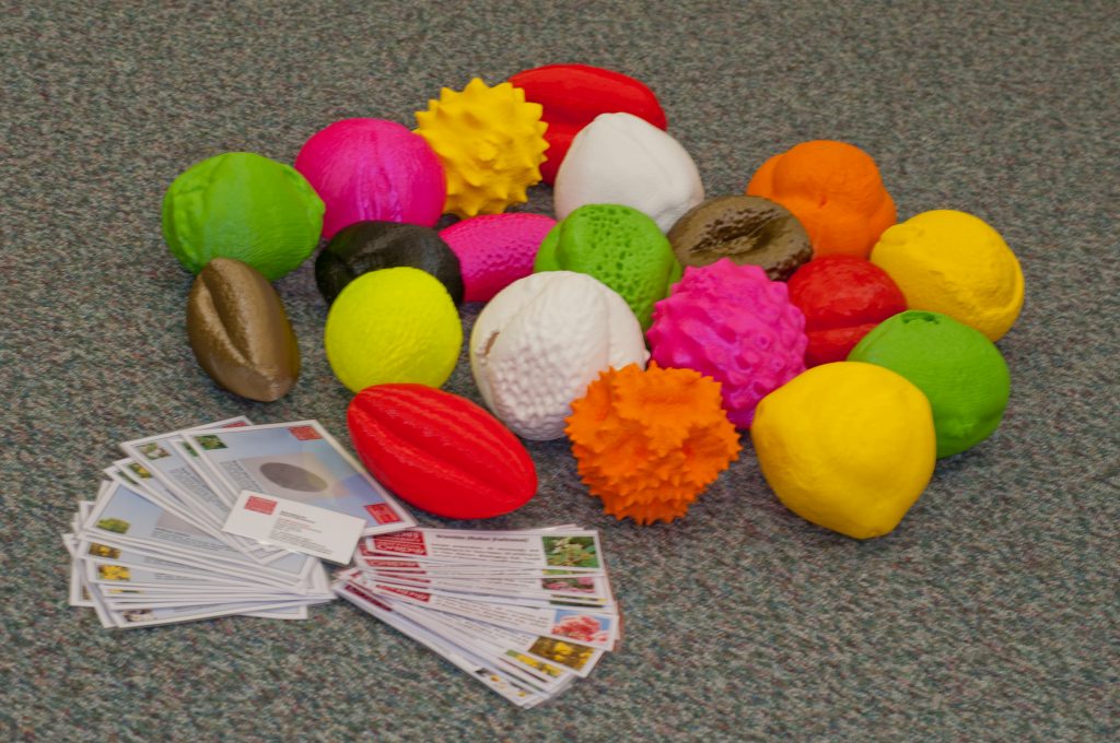
Recently, we were approached by the National Botanic Gardens of Wales (NBGW) to generate 3D models of twenty different species of pollen grains identified in honey by Dr Natasha De Vere’s research group for their science outreach and engagement programme. Natasha is the head of science at the NBGW and is using cutting edge DNA bar coding technology to understand pollinator foraging preferences. This research is providing amazing insights into the selective range of plant species that important pollinating insects such as bees visit when foraging (you can read more about this fascinating work here and in the reference below).
To generate the 3D prints we first needed pollen samples from each of the respective plant species – you’d be forgiven if you thought the National Botanic Gardens could provide these ‘off the shelf’ : ) Coming from a zoological background, my botany field skills are best described as rudimentary. So, equipped with my smartphone, a plant identifier app that I downloaded from the Google Play store, and some zip-lock sample bags, I embarked upon a ‘Pokemon-go style’ palynological quest (‘gotta catch ’em all’) that took me to the local parks, woodlands, river embankments, country lanes and coastlines and even garden centres of South Wales.
After some effort, I managed to identify and collect all of the pollen species on the wish list. I then began to image representative grains from each species using the Hub’s Zeiss LSM880 Airyscan confocal microscope. Individual grains were optically sectioned through their volume, 3D reconstructed and then output in a file format for 3D printing on our Ultimaker 3D printer (method described in reference below).
The finished 3D pollen models are shown in the photograph above – each model is approximately 15cm in diameter (i.e., enlarged by a factor of approximately x400 relative to the original pollen grain). The models will be on display at the Growing the Future stand at this year’s Royal Welsh show (20-23rd July, 2019) and also at the Pollinator festival at the National Botanical Gardens of Wales (24-26th August, 2019).
AJH
Further Reading
Hawkins, J., de Vere, N., Griffith, A., Ford, C.R., Allainguillaume, J., Hegarty, M.J., Baillie, L., Adams-Groom, B. (2015) Using DNA metabarcoding to identify the floral composition of honey: a new tool for investigating honey bee foraging preferences. PLoS ONE10 (8): e0134735. https://doi.org/10.1371/journal.pone.0134735.
Sample collection and preparation, confocal microscopy, 3D reconstruction and file conversions by Dr Tony Hayes; 3D printing by Dr Pete Watson; Photography by Marc Isaacs.
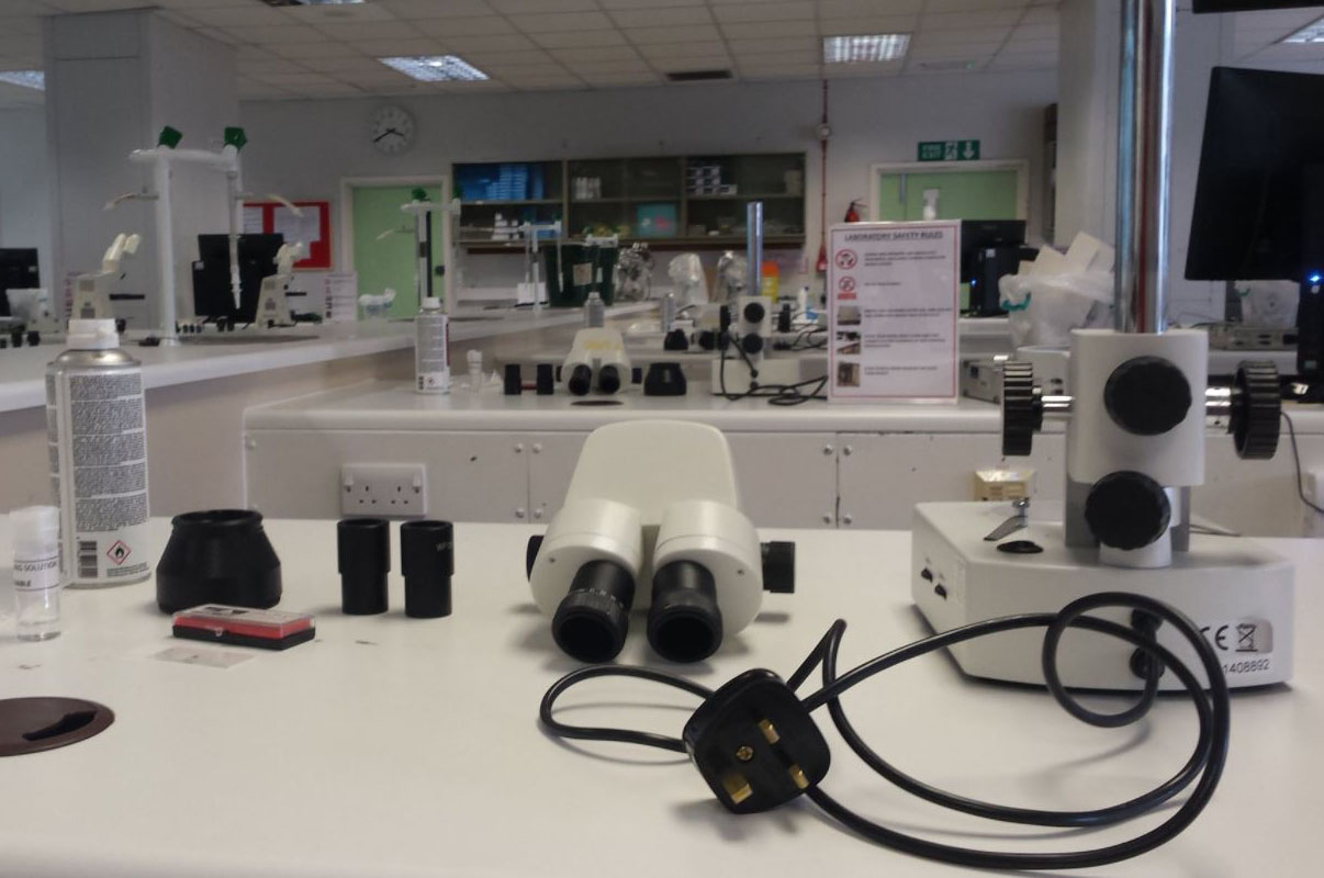
Above: Some of the class microscopes in various states of dismantlement.
It goes without saying, to get the very best out of a microscope you need to know how to optimise and maintain its performance. That said, you’d be surprised just how many microscopists don’t know how to properly set up and maintain their microscopes.
Recently, we run our first microscope maintenance course as part of Cardiff University’s Continuous Professional Development (CPD) programme. We can’t tell you who it was for; but suffice to say, they use microscopes a lot in their work. The two day course covered the basics of light microscopy and the procedures necessary to keep a microscope squeaky- clean and correctly aligned. The practical element of the course saw delegates clean, rebuild and align both upright widefield and stereo-zoom microscopes.
Pleasingly, the course was well-received with very good to excellent feedback. Thanks to all who participated on the two busy but enjoyable days. Thanks must alsogo to our undergrad students for soiling and mis-aligning the microscopes ahead of the course – they did a far better job than we ever could ; )
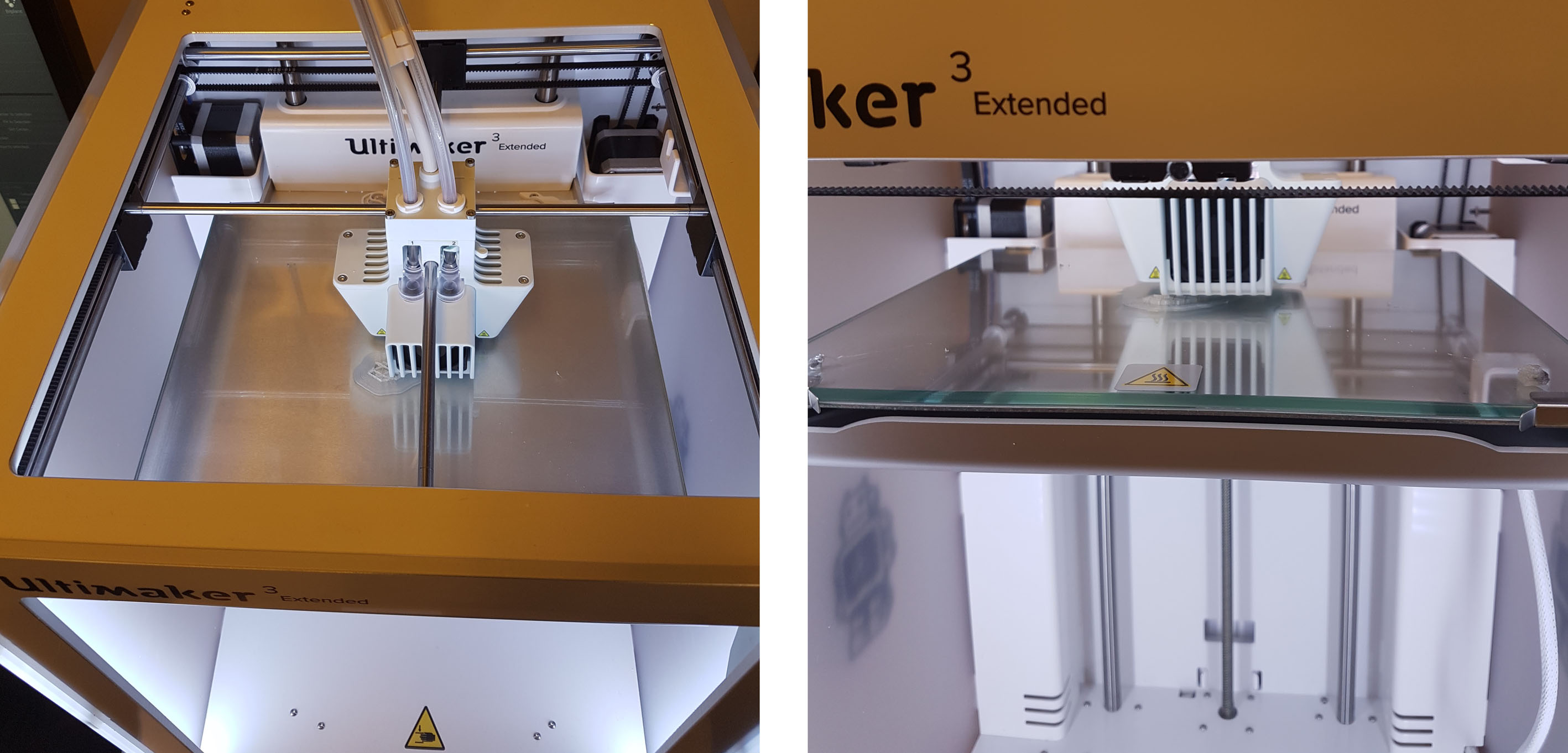
Above: The new Ultimaker ‘Extended’ 3D printer building a stage insert for a novel microscope system.
The Bioimaging Research Hub has recently purchased an Ultimaker 3 ‘Extended’ professional 3D printer. The printer will allow users to 3D print bespoke pieces of scientific equipment or generate scale models of microscopic samples for use in their research as well as for teaching, outreach and engagement activities (examples can be found here andhere).
The 3D printer utilises FDM (Fused Deposition Modelling) printing technology and has a range of advanced features allowing the fabrication of professional quality, high resolution 3D prints. The printer can print in two different colours or a single colour in addition to a dissolvable PVA support scaffold, thus allowing complex overhanging structures to be printed at high fidelity whilst significantly reducing finishing time.
The printer has a large build volume (215 x 215 x 300mm), supports a range of materials (nylon, PLA, ABS, CPE and PVA) and has a print resolution of 20-200 microns. The printer is wi-fi enabled with an internal webcam so that users can remotely monitor the progress of their 3D prints.
Further details of the equipment are available through our equipment database.
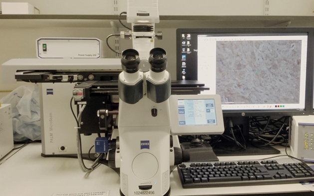
Above: ‘Cutting-edge’ equipment: the Zeiss PALM MicroBeam laser micro-dissector located at the European Cancer Stem Cell Research Institute (ECSCRI)
A Zeiss PALM MicroBeam laser micro-dissector system is now available as a spoke of the Bioimaging Research Hub. The equipment, which is located at the European Cancer Stem Cell Research Institute (ECSCRI), allows isolation of DNA, RNA and protein from laser micro-dissected samples from both histological sections (paraffin wax or cryo) and live cells. Further details of the system are available through our equipment database. All enquiries for this system should be directed towards Mr Mark Bishop.
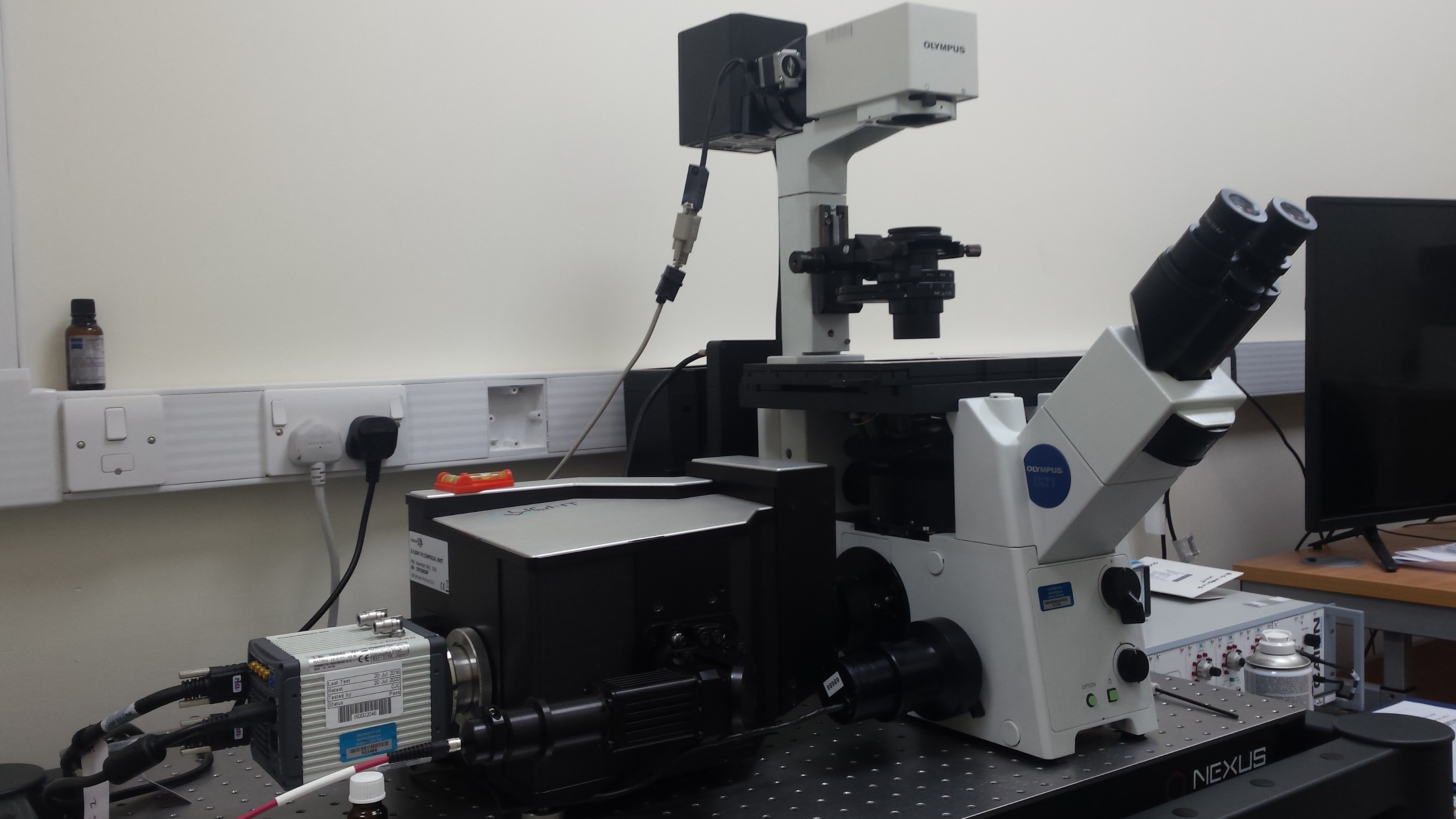
The old electron microscopy darkroom (BIOSI 2; E/0.05) within the Bioimaging Research Hub has recently been refurbished as a live cell imaging suite via generous support from BIOSI. It now houses a spinning disc confocal system for fast, live cell imaging applications. The system is based around an Olympus IX71 inverted microscope, kindly provided by Dr Pete Watson, which has been upgraded, via ISSF funding, with a Crest Optics X-Light V2 confocal head, a Cairn Research tri-line laser bank (405nm, 488nm, 561nm) and a Hamamatsu ORCA Flash 4 sCMOS digital camera with M-View Gemini image splitter. The system is fully integrated via Molecular Devices MetaMorph software and boasts a 40″ 4k display. The system will expand the Hub’s imaging toolbox, enabling high speed, multi-position, multi-colour 3D/4D image acquisition. Support systems for live cell imaging (i.e. gas and incubation) are also available within the facility. Further details of this system are available through our equipment database.