EQUIPMENT: New State-of-the-Art Confocal Microscope with Super-Resolution Capability.

DSC_0459

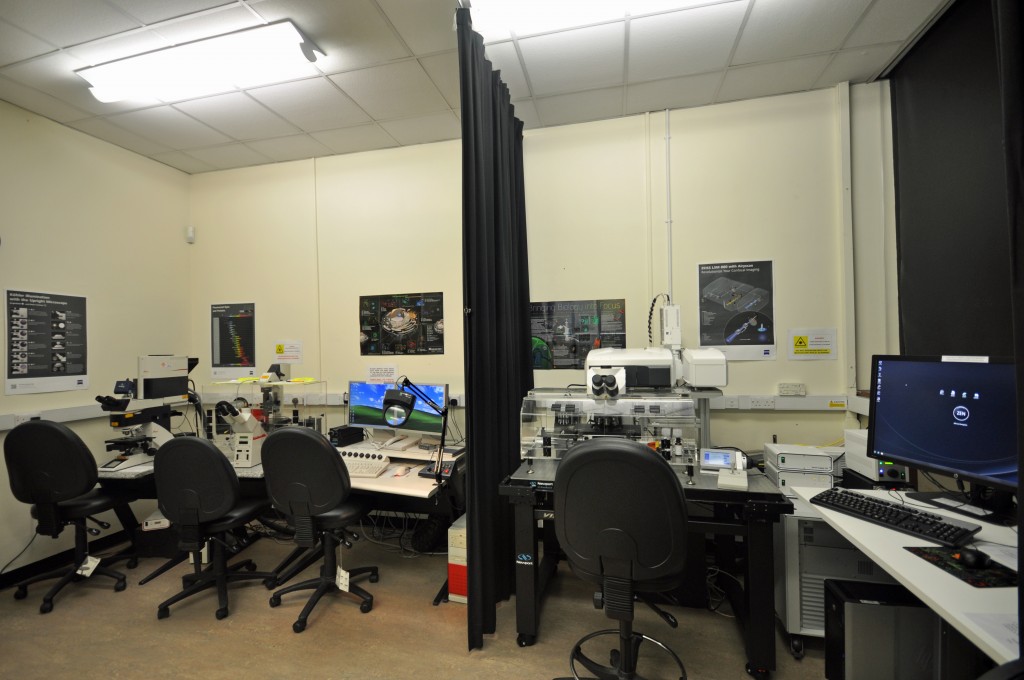
Image: The old and the new: The new Zeiss LSM 880 Airyscan confocal system (right) adjacent to the old Leica TCS SP2 confocal (left) (BIOSI 2; E/0.03)

The BIOSI Bioimaging Research Hub has recently expanded its imaging toolbox with a new, state-of-the-art confocal microscope system, that was purchased through generous funding by the Research Infrastructure Fund (Lead Applicant: Dr Walter Dewitte). The system, a top-of-the-range Zeiss LSM 880 upright confocal microscope, features the advanced Airyscan super-resolution detection module which provides a 1.7x gain in resolution in all three dimensions compared to conventional confocal optics. The system also supports advanced fluorescence techniques including FCS (fluorescence correlation microscopy) and FLIM (fluorescence lifetime imaging (FLIM) – the FLIM module will be installed during the first week of December. Further details here.

AJH

Find out more:

NEWS: Newly-refurbished microscopy suite.

DSC_0456

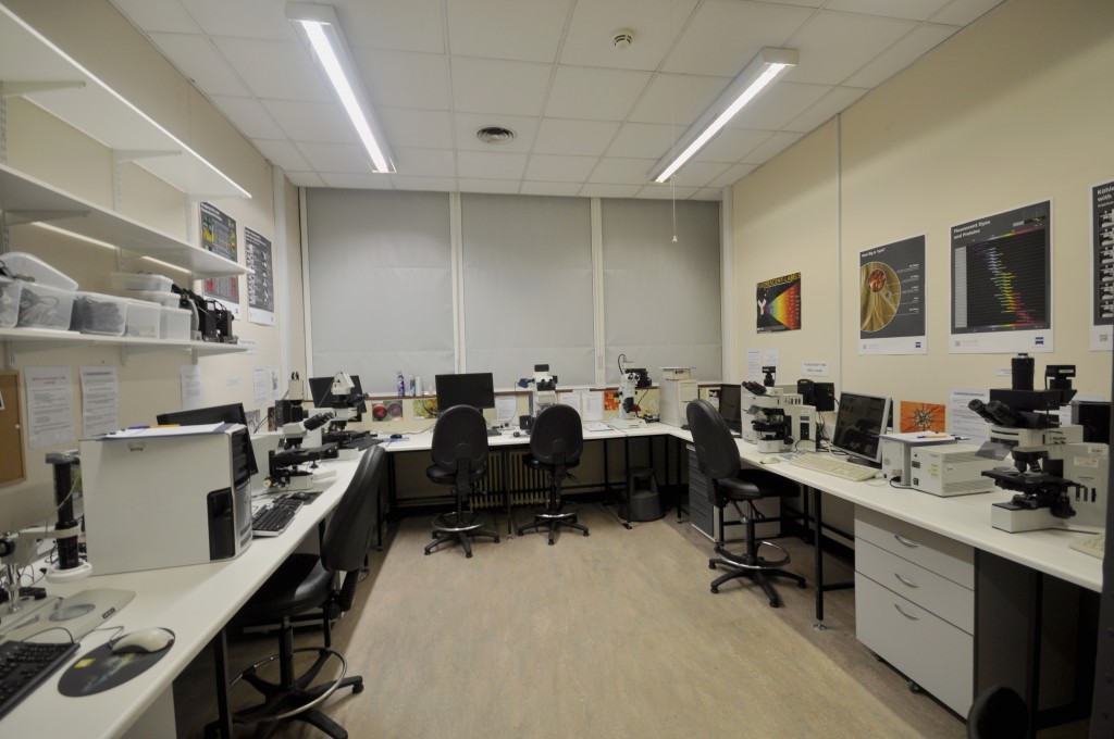
Image: The newly-refurbished microscopy suite (BIOSI 2; E/0.04).

The old SEM room (BIOSI 2; E/0.04) has now been refurbished as a dedicated widefield microscopy suite via School support. The refurbishments include new electrical and internet connections, benching, secondary glazing and black-out blinds. The suite hosts a broad range of modern transmitted light and epifluoresence systems including (from left to right):

All of the systems are available for use with or without technical support and are suitable for a wide variety of research purposes, including student projects.


INTRODUCTION.

This is a short post to outline what you will find in each of the sections within this blog.

  • Features contains information on recent events within the Bioimaging facility (e.g. new equipment, interesting research applications etc). Posts will be labelled either NEWS for general news items, IN-FOCUS for particular highlights of applications within the Unit or EQUIPMENT for new pieces of equipment in the unit.
  • Resources highlights some of the microscopical imaging techniques that are currently available and how these may benefit your research.
  • Gallery showcases a selection of the microscopical images produced within the facility.
  • Publications provides a list of all known publications (with linkouts) arising from research conducted within the facility.
  • FAQs contains answers to Frequently Asked Questions, fluorochrome data and other technical miscellany.

We hope that you find the information contained within this blog useful, or at least interesting, and look forward to your comments,

The Bioimaging Unit