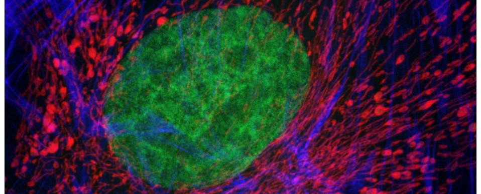
The Bioimaging Hub has recently completed work in digitising the School’s extensive histopathology slide repository. Over 400 histological sections, encompassing both normal and pathological tissues, were painstakingly scanned and digitised in high resolution using the facilities Objective Imaging Surveyor slide scanning system. The datasets, totalling 4TB, have been converted into the Zoomify .ziff image file format to enable easy and rapid on-line browsing, zooming and navigation (similar to that of Google Earth) and calibrated to allow feature measurement. The image files have been linked, via thumbnails, to a database that captures all relevant metadata for each histological section (filename, tissue type, organ system, species, section plane, histological stain, section ID, supplier, objective magnification etc) to facilitate easy sorting and data retrieval. The database is currently set up on a basic Linux server within the facility; however, to cope with concurrent file access by large numbers of up to 150 students, it will require a permanent home on a dedicated server within the School. With further development, the resource promises to have fantastic potential for teaching, research and public engagement within BIOSI. Thanks to all concerned who have taken the project this far…
AJH
Find out more:
- Ghaznavi et al (2013) Digital imaging in pathology: whole slide imaging and beyond. Annu Rev Pathol Mech Dis 8: 331-359.
- Hamilton et al (2012) Virtual microscopy and digital pathology in training and education. APMIS 120: 305-315.

