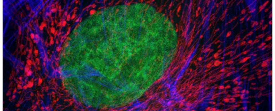
So, with the advent of spring, we thought it was high time we dusted down our YouTube channel and gave it an overhaul. As well as introducing lots of nice new image content from some of our latest 3D imaging systems, we felt that the channel would have a greater sense of purpose if we were to develop it into an educational resource for microscopy and bioimaging, with obvious relevance for remote learning (i.e., perfect in our current circumstances). Consequently, we have started to collate the most useful and relevant of YouTube’s microscopy-related content (webinars, tutorials, demonstrations etc), ranging from the basic principles of light microscopy to cutting-edge fluorescence-based nanoscopy techniques such as FLIM, so that they are all placed under one roof for your convenience : )
One of the things we hoped to provide our userbase was a series of video tutorials for the hub’s many microscope systems. Whilst there’s a great deal of useful training material on YouTube, in the main, it tends to be aimed at many of the high-end, turn-key imaging systems. Furthermore, not all microscopes are created equal, they each have their own peculiarities which reflect their intended function and most ‘evolve’ over time, through upgrades, to accomodate the vagaries of research. So, with this in mind, we have started to create our very own bespoke training videos for each of the hub’s microscope systems (example here).
The new training videos will supplement the standard operating procedures (SOPs) we have written for all of our imaging equipment and should provide an invaluable resource for user training and e-learning. As such, they will be embedded within the appropriate sections of the hub’s SOP repository (read more here). The online video content and their associated SOPs will be viewable at the click of a mouse button via desktop shortcuts on all of our microscope-associated PCs allowing easy access during instrument operation.
If you have time on your hands, then please pop over to YouTube and take a look at how our channel is developing (link here). It’s still work in progress but, as I say, it has the potential to be an extremely useful resource; not only for hub users, but anyone with a passing interest in microscopy and bioimaging. Constructive feedback is welcomed.
AJH.
