The Bioimaging research hub does a nice sideline 3D printing scale replicas of biological samples for use in science engagement and teaching. These can be made from the smallest of microscopic samples imaged via optical sectioning microscopy (i.e., confocal or lightsheet) or from large anatomical samples imaged via 3D photogrammetry or from 3D scanning techniques. I’ve posted a few blogs on this site in the past describing 3D pollen models that we’ve made for various research groups within Cardiff University (e.g. for the ‘Footprints in time’ and ‘PharmaBees’ projects) and for a growing number of external organisations within the UK and abroad (e.g. the Met office, the Smithsonian institute etc).
Recently, we were approached by the National Botanic Gardens of Wales (NBGW) to generate 3D models of twenty different species of pollen grains identified in honey by Dr Natasha De Vere’s research group for their science outreach and engagement programme. Natasha is the head of science at the NBGW and is using cutting edge DNA bar coding technology to understand pollinator foraging preferences. This research is providing amazing insights into the selective range of plant species that important pollinating insects such as bees visit when foraging (you can read more about this fascinating work here and in the reference below).
To generate the 3D prints we first needed pollen samples from each of the respective plant species – you’d be forgiven if you thought the National Botanic Gardens could provide these ‘off the shelf’ : ) Coming from a zoological background, my botany field skills are best described as rudimentary. So, equipped with my smartphone, a plant identifier app that I downloaded from the Google Play store, and some zip-lock sample bags, I embarked upon a ‘Pokemon-go style’ palynological quest (‘gotta catch ’em all’) that took me to the local parks, woodlands, river embankments, country lanes and coastlines and even garden centres of South Wales.
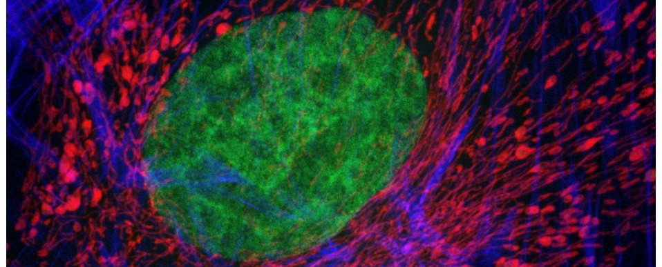
After some effort, I managed to identify and collect all of the pollen species on the wish list. I then began to image representative grains from each species using the Hub’s Zeiss LSM880 Airyscan confocal microscope. Individual grains were optically sectioned through their volume, 3D reconstructed and then output in a file format for 3D printing on our Ultimaker 3D printer (method described in reference below).
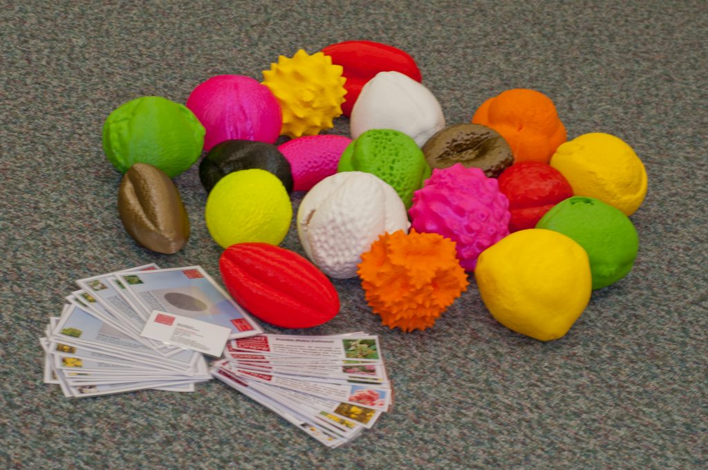
The finished 3D pollen models are shown in the photograph above – each model is approximately 15cm in diameter (i.e., enlarged by a factor of approximately x400 relative to the original pollen grain). The models will be on display at the Growing the Future stand at this year’s Royal Welsh show (20-23rd July, 2019) and also at the Pollinator festival at the National Botanical Gardens of Wales (24-26th August, 2019).
AJH
Further Reading
Hawkins, J., de Vere, N., Griffith, A., Ford, C.R., Allainguillaume, J., Hegarty, M.J., Baillie, L., Adams-Groom, B. (2015) Using DNA metabarcoding to identify the floral composition of honey: a new tool for investigating honey bee foraging preferences. PLoS ONE 10 (8): e0134735. https://doi.org/10.1371/journal.pone.0134735.
Perry, I., Szeto, J-Y., Isaacs, M.D., Gealy, E.C., Rose, R., Scofield, S., Watson, P.D., Hayes, A.J. (2017) Production of 3D printed scale models from microscope volume datasets for use in STEM education. EMS Engineering Science Journal. 1 (1): 002.
Contributors
Sample collection and preparation, confocal microscopy, 3D reconstruction and file conversions by Dr Tony Hayes; 3D printing by Dr Pete Watson; Photography by Marc Isaacs.
