Skip to content

Bioimaging Hub News

News from the Bioimaging Research Hub in the School of Biosciences, Cardiff University.

  • Home
  • Features
    • CORE EQUIPMENT: New Spinning Disc Confocal System for Live Cell Imaging.
    • SPOKE EQUIPMENT: Zeiss PALM MicroBeam Laser Micro-dissector.
    • IN FOCUS: Cutting Through the Fog: Reducing Background Autofluorescence in Microscopy.
    • IN-FOCUS: Better To Burn Bright Than To Fade Away: Reducing Photo-bleaching in Fluorescence Microscopy.
    • IN-FOCUS: Development of a 3D Printed Pollen Reference Collection.
    • IN-FOCUS: Microscopy on the move. A round-up of the best microscopy apps for mobile devices.
    • IN-FOCUS: Bigging It Up: 3D Printing to Change the Shape of Microscopy.
    • IN-FOCUS: Microscopy and Analysis Journal: A Useful Resource for Microscopists.
    • CORE EQUIPMENT: PicoQuant FLIM upgrade for the Zeiss LSM880 Airyscan Confocal Microscope.
    • NEWS: Science, Technology, Engineering & Mathematics (STEM) Conference, 2015.
    • IN-FOCUS: Imaging on a Budget? A Round-up of the Best Free Imaging Software on the Web.
    • IN-FOCUS: De-boning the Zebrafish: unpicking skeletogenesis under the microscope.
    • IN-FOCUS: Gumming up the works: oral biofilms under the microscope.
    • IN-FOCUS: Shedding light on new fluors: development of novel, metal-based probes for bioimaging.
    • NEWS: Naomi’s nightmares of nature.
    • IN-FOCUS: Heavy metal worms under the spotlight.
    • SPOKE EQUIPMENT: New Multi-Spectral In Vivo Imaging system.
  • Gallery
  • Publications
  • Covers
  • FAQs
  • 3D Pollen Library
Bioimaging Hub News

Fluorescence Lifetime IMaging (FLIM)

What is Fluorescence Lifetime IMaging (FLIM)  and how can it help me in my research?

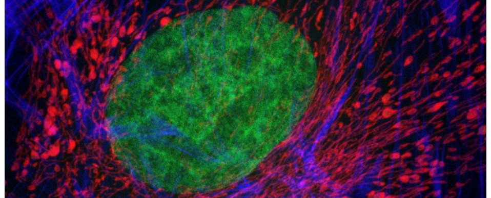
Fluorescence-lifetime imaging is a microscopical technique for producing an image based on the differences in the exponential decay rate of the fluorescence from a sample.
Find out more:
  • Leica Science Lab
  • Nikon Microscopy U
  • Olympus Microscopy Resource Centre
Our FLIM system:
  • Zeiss LSM 880 confocal microscope

Search this blog

Recent features:

  • IN FOCUS: Masson’s trichrome – but which one?
  • IN FOCUS: Breaking the Mould: Supersizing Pollen Grains as Gigantic Public Art Sculptures.
  • IN FOCUS: The ‘3D Pollen Library’: An Update
  • IN FOCUS: Making the Most of Your Microscope.
  • IN FOCUS: AR Palynology: Probing the Reality of Nature/Nature of Reality.
  • NEWS: New BIOSI Live Cell Imaging Spoke
  • CORE EQUIPMENT: New Zeiss Celldiscoverer 7 system
  • NEWS: Updated Covid Rules: Resumption of Hands-on Support and Training.
  • IN-FOCUS: Brushing Up On Your Background Knowledge.
  • SPOKE EQUIPMENT: Olympus VS200 High Throughput Slide Scanning Microscope.
  • NEWS: Reopening of the Bioimaging Research Hub: New COVID 19-Security Measures.
  • IN FOCUS: Bioimaging Hub’s YouTube Channel – Rebooted.
  • NEWS: State of the Art: An Update.
  • IN FOCUS: Plastic Fantastic – Making Pollen Models for The National Botanic Garden of Wales.
  • CORE EQUIPMENT: Widefield Microscope Upgrades.
  • IN FOCUS: Winging It In Paleobiology: Strange Tails from a Strange Time.
  • IN FOCUS: Immersive Microscopy – 3D Visualisation and Manipulation of Microscopic Samples Through Virtual Reality.
  • IN FOCUS: Standard Operation Procedures (SOP) Repository.
  • IN FOCUS: Imaging Cleared Tissues by Lightsheet Microscopy.
  • IN-FOCUS: Making Imaris a Bit(plane) Faster
  • SPOKE EQUIPMENT: X-Clarity Tissue Clearing System.
Proudly powered by WordPress