![Fluorescence image of neurons showing four distinct coloured stains](http://blogs.cardiff.ac.uk/bioimaging/wp-content/uploads/sites/492/2015/11/neurones_001-300x260.jpg)
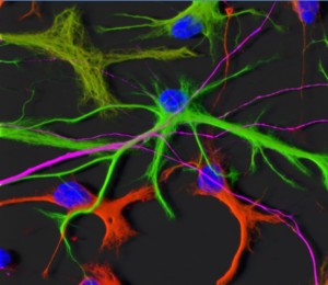
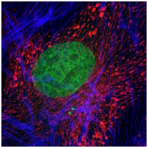
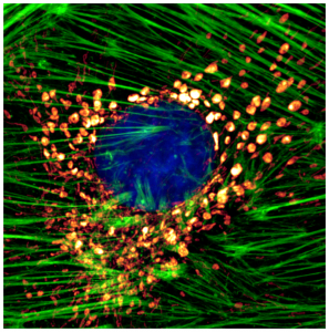
“Optimising Brain Cell Imaging: Gray Matters”
![3D surface-rendered model of appendage (endopod of maxilliped) of Chinese mitten crab larva](http://blogs.cardiff.ac.uk/bioimaging/wp-content/uploads/sites/492/2016/01/Picture10-300x282.png)
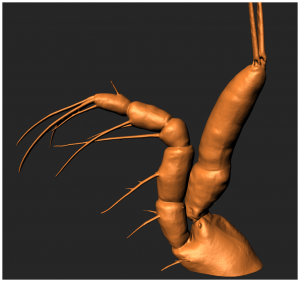
“Imaging The Mitten Crab: Working Hand In Glove With The Natural History Museum” (Image 1 of 2)
![abdominal segment of a mitten crab larva](http://blogs.cardiff.ac.uk/bioimaging/wp-content/uploads/sites/492/2016/01/mitten-crab-300x276.png)
“Imaging The Mitten Crab: Working Hand In Glove With The Natural History Museum” (Image 2 of 2)
![image of the compound eye of a common fly](http://blogs.cardiff.ac.uk/bioimaging/wp-content/uploads/sites/492/2016/01/Picture8-300x285.jpg)
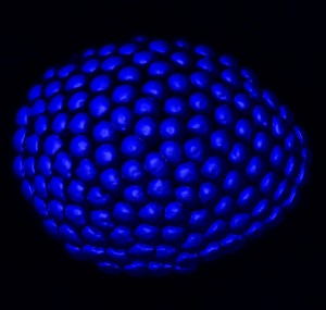
“Compound Eye of Garden Ant”
![two endothelial cells showing a region of binding between them](http://blogs.cardiff.ac.uk/bioimaging/wp-content/uploads/sites/492/2016/01/Picture6-297x300.jpg)
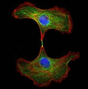
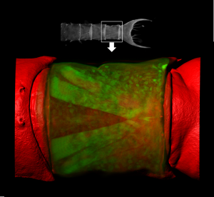
“The Secret Lives Of Cells (4096 Shades Of Gray)”
![an endothelial cell with nuclear contents showing a smiling face](http://blogs.cardiff.ac.uk/bioimaging/wp-content/uploads/sites/492/2016/01/Picture4-300x300.jpg)
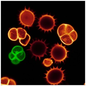
“Have A Nice Day ; )”
![several grains of pollen](http://blogs.cardiff.ac.uk/bioimaging/wp-content/uploads/sites/492/2016/01/Picture3-300x300.jpg)
“Nothing To Be Sneezed At”
![A renal corpuscle](http://blogs.cardiff.ac.uk/bioimaging/wp-content/uploads/sites/492/2016/01/Picture9-300x300.png)
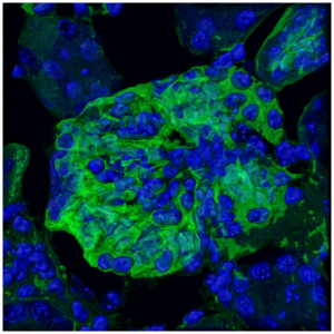
“Encapsulating Kidney Function”
![Endothelial cell showing mitochondria and the cytoskeleton](http://blogs.cardiff.ac.uk/bioimaging/wp-content/uploads/sites/492/2016/01/Picture5-297x300.png)
“Shedding Light On Cellular Function”
![A mosaic image of a zebrafish using various image processing methods](http://blogs.cardiff.ac.uk/bioimaging/wp-content/uploads/sites/492/2016/01/Picture2-300x300.jpg)
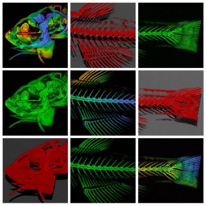
“De-boning The Zebrafish: Unpicking Skeletogenesis Under The Microscope”
![Candida albicans biofilm.](http://blogs.cardiff.ac.uk/bioimaging/wp-content/uploads/sites/492/2015/11/Candida-biofilm-300x300.jpg)
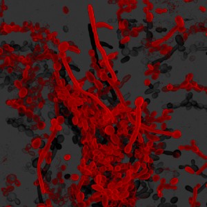
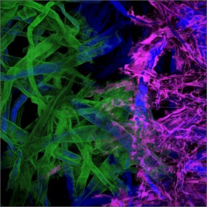
“Gumming Up The Works: Oral Biofilms Under The Microscope”
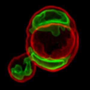
![A yeast cell with a bud that has cell contents being transferred into it](http://blogs.cardiff.ac.uk/bioimaging/wp-content/uploads/sites/492/2016/01/5516988939_b345aa2fb7_o-300x300.jpg)
“Blooming Yeast!”
![Serial number of a ten pound note](http://blogs.cardiff.ac.uk/bioimaging/wp-content/uploads/sites/492/2016/01/5516986793_2fec8c5d33_o1-300x300.jpg)
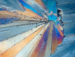
“The Colour Of Money”
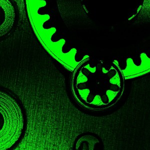
![Internal workings of a watch mechanism.](http://blogs.cardiff.ac.uk/bioimaging/wp-content/uploads/sites/492/2016/01/5516985939_fc26565bc0_o1-300x300.jpg)
“Time To Reflect”
![Tissue paper coloured with highlighter pens](http://blogs.cardiff.ac.uk/bioimaging/wp-content/uploads/sites/492/2016/01/5517575926_b439612eaf_o1-300x300.jpg)
“Putting Pen To Paper”
![polarised light image of sugar crystals](http://blogs.cardiff.ac.uk/bioimaging/wp-content/uploads/sites/492/2016/01/5517575816_e6bb3933a5_o-300x226.jpg)
“All Sweetness And Light”
WORK IN PROGRESS: A further selection of images produced within the Unit can be viewed on our Flickr and YouTube pages.