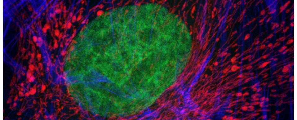
What is multiphoton microscopy and how can it help me in my research?
Multiphoton microscopy is a form of laser-scanning microscopy that uses non-linear optics to achieve optical sectioning of fluorescent samples. In multiphoton microscopy, a pulsed, long-wavelength, low energy (typically infra-red) laser is used to excite fluorescence at the plane of focus within a sample. When the photon density is sufficiently high (i.e. two molecules arriving within 1 femtosecond of each other), photons of lesser energy together cause an excitation normally produced by the absorption of a single photon of higher energy. It thus differs from traditional fluorescence imaging, in which the excitation wavelength is shorter than the emission wavelength.
The multiphoton microscope shares many similarities with the confocal microscope (i.e. raster scanning and optical sectioning) but also offers some advantages: (1) multiphoton microscopy only excites fluorescence at the plane of focus, thus does not cause photobleaching or phototoxicity outside this plane; (2) it does not require a pinhole aperture for optical sectioning, thus no fluorescent light is rejected during image acquisition; and (3) the longer wavelength excitation lasers of multiphoton systems have deeper tissue penetration (up to 1mm) and cause less damage than short-wavelength lasers, thus cells/tissues may be observed for longer periods with fewer toxic effects.
