
Imaging The Mitten Crab: Working Hand In Glove With The Natural History Museum
“Imaging The Mitten Crab: Working Hand In Glove With The Natural History Museum” (Image 2 of 2)
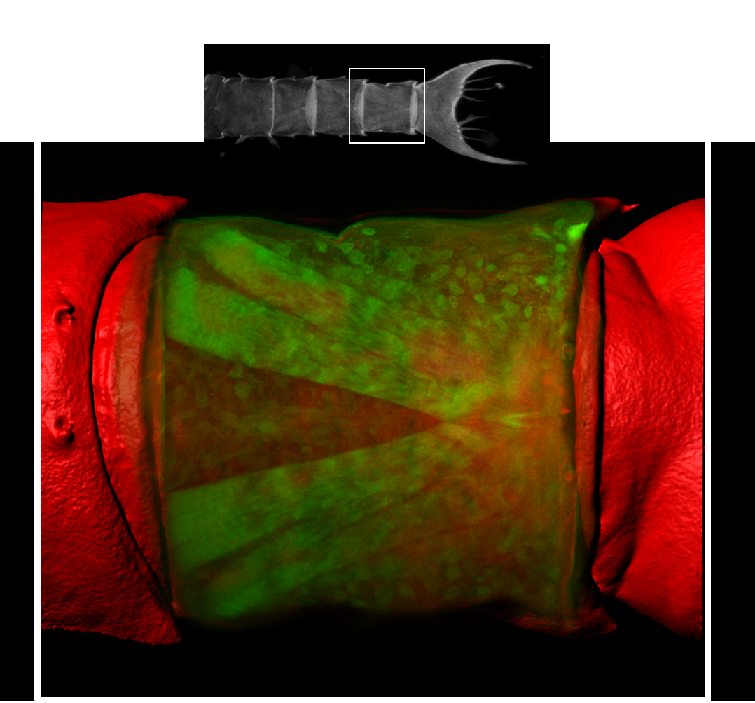
Confocal reconstruction of an abdominal segment within the tail region of a Chinese Mitten crab larva (Eriocheir sinesis). The exoskeleton has been surface-modelled (red) whilst the underlying musculature is shown in green. Image produced by Dr Tony Hayes on a Zeiss LSM 880 confocal system. Sample processed by Seyit Ali Kamanli (Natural History Museum). Click image to enlarge.
Further information on this application can be found in the following IN-FOCUS post: Imaging the Mitten crab: working hand in glove with the Natural History Museum