
Imaging The Mitten Crab: Working Hand In Glove With The Natural History Museum (Image 1 of 2)
“Imaging The Mitten Crab: Working Hand In Glove With The Natural History Museum (Image 1 of 2)”.
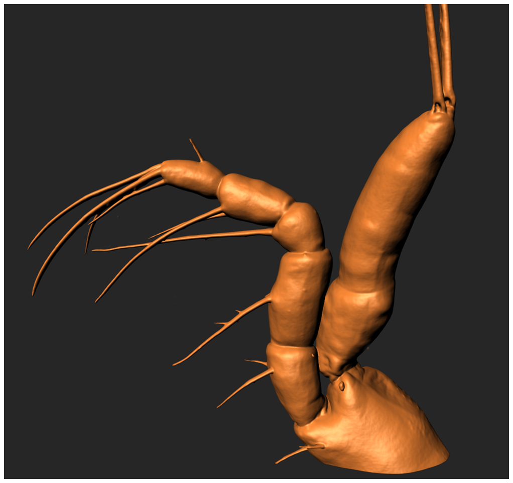
Confocal surface reconstruction showing the endopod of a maxilliped taken from a Chinese Mitten crab larva (Eriocheir sinesis). Image produced by Dr Tony Hayes on a Zeiss LSM 880 confocal system. Sample processed by Seyit Ali Kamanli (Natural History Museum). Click image to enlarge.
Find out more here.