Minimally invasive diagnosis and prognostic markers for mast cell tumours in companion animals
18 April 2026
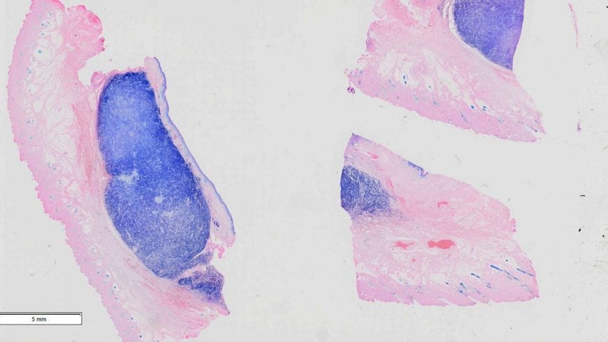
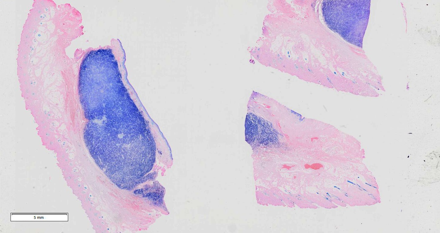
Mast cell tumours are a common cancer in companion animals, particularly dogs, but it can be difficult to decide which treatment to use on them.
This research focuses on developing new approaches to understand these tumours, with the aim of supporting earlier diagnosis, less invasive testing, better monitoring and improved care for pets.
In National Pet Month, we’re looking at how Cardiff University research is helping us care for and treat our pets.
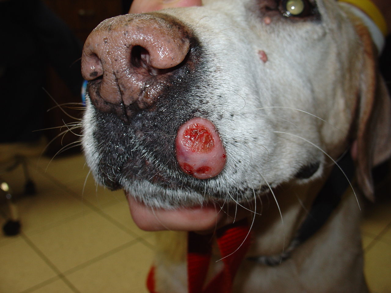
Impact funding from the Biotechnology and Biological Sciences Research Council, managed by Cardiff University, has enabled new approaches to detect and monitor mast cell tumours.
Led by Professor Matt Smalley, this work is developing minimally invasive methods to support earlier diagnosis and better outcomes for pets.
Researchers developed a minimally invasive molecular approach to detect tumour specific changes in dogs using different sample types, including blood samples, reducing the need for invasive biopsies.
The study demonstrated accurate detection of tumour associated mutations in multiple sample types, offering flexibility in testing to decide on the best course of tumour treatment.
Impact Acceleration Account (IAA) funding has enabled development of new diagnostic approaches and supported collaboration with partners including Finn Pathologists and industry suppliers.
This work has generated the data needed to move towards clinical application and commercialisation, positioning the research for follow on funding, licensing opportunities and future use in veterinary practice.
- AHRC
- BBSRC
- Cardiff Business School
- Case studies
- EPSRC
- ESRC
- Institutional Collaboration
- MRC
- Public Engagement
- School of Biosciences
- School of Computational and Mathematical Sciences
- School of Dentistry
- School of Engineering
- School of English, Communication and Philosophy
- School of Geography and Planning
- School of History, Archaeology and Religion
- School of Journalism, Media and Culture
- School of Law and Politics
- School of Medicine
- School of Optometry and Vision Sciences
- School of Psychology
- Welsh School of Architecture
- Eight Voices in Darkness recognised for sharing lived experiences of sight loss and mental health
- How can research become more inclusive for people who have difficulty giving informed consent?
- How can medical imaging software developed in Cardiff reach more researchers, clinicians and healthcare innovators?
- What does it take to make secondhand economies more sustainable, accessible and socially connected?
- What happens when retailers cannot trust their stock records?